


 Медицина
Похожие презентации:


Autopsy report Patient N, 76 уears
1.
Autopsy reportPatient N, 76 уears
2.
SUMMARY OF CLINICAL HISTORY:• Patient N, 67 years old, suffered from ovarian cancer.
• She underwent bilateral laparoscopic adnexectomy.
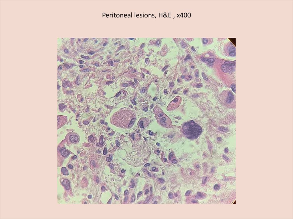
• Microscopic and IHC examination : undifferentiated endometrioid
carcinoma, CEA 4 +, HER2neu-negative
• Received polychemotherapy since July 2020.
• In September her condition significantly deteriorated.
• Was hospitalized
• Was diagnosed with carcinomatosis of the peritoneum. Ascites.
Metastasis in S 10 of the right lung.
3.
EXTERNAL EXAMINATION:• The body of 77 year old women, well developed,
well nourished.
• Skin: pale gray
•On the anterior surface of the chest and lower third
of the lower limbs - multiple petechial hemorrhages
•In the projection of the thyroid cartilage there is an
old scar 4 cm long, corresponding to the scar after
hemithyroidectomy
•In the epigastric, umbilical and left hypochondriac
regions there are scars up to 2 cm in diameter,
corresponding to the laparoscopy
•cadaveric spots situated on the back of the body,
pale purple, turn pale when pressure
4.
INTERNAL EXAMINATION5.
Heart•Size – 11х9х5 cm
•Weight– 336 g
•Wall thickness of the left ventricle 1.5 cm, right 0.3
cm.
• Epicardium and pericardium are smooth, shiny,
with moderate adipose tissue deposition
• Heart valves are smooth, shiny, perimeter within
normal limits
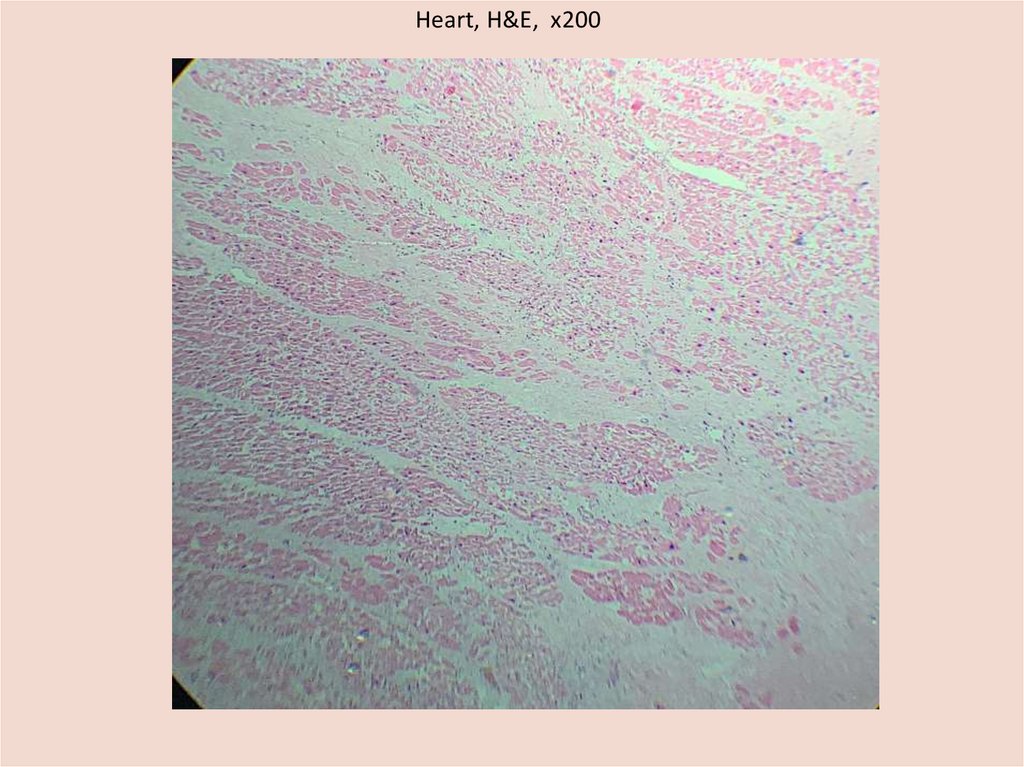
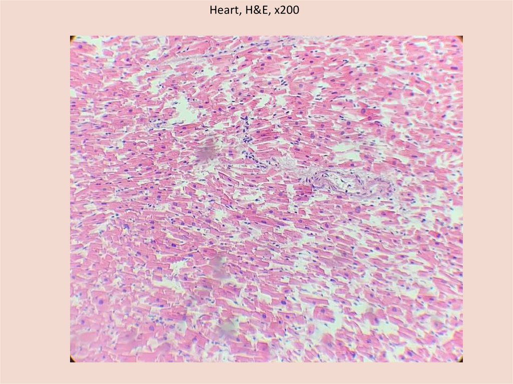
•Myocardium is pale brown, foci of uneven blood
supply. In the lateral wall of the left ventricle, a focus
of irregular shape, dense, whitish colour, size -4x3x1
cm
6.
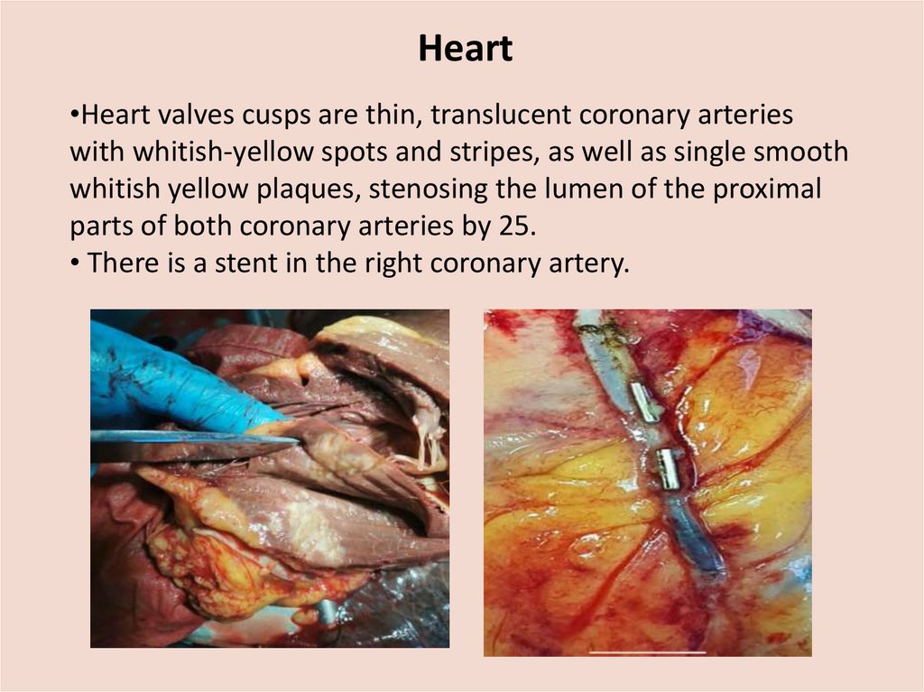
Heart•Heart valves cusps are thin, translucent coronary arteries
with whitish-yellow spots and stripes, as well as single smooth
whitish yellow plaques, stenosing the lumen of the proximal
parts of both coronary arteries by 25.
• There is a stent in the right coronary artery.
7.
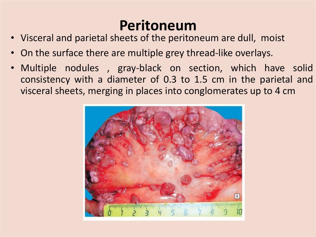
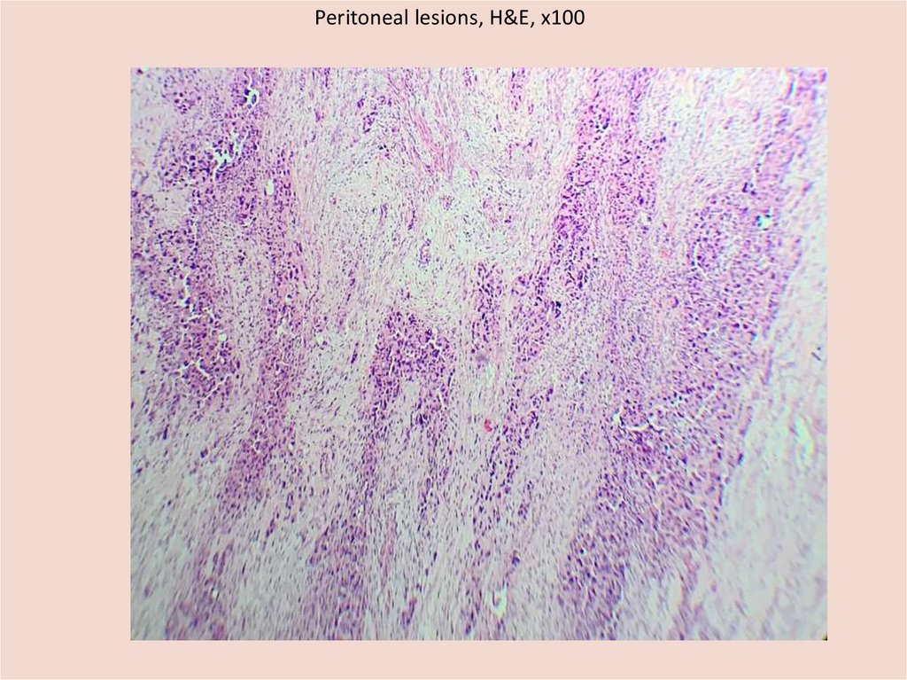
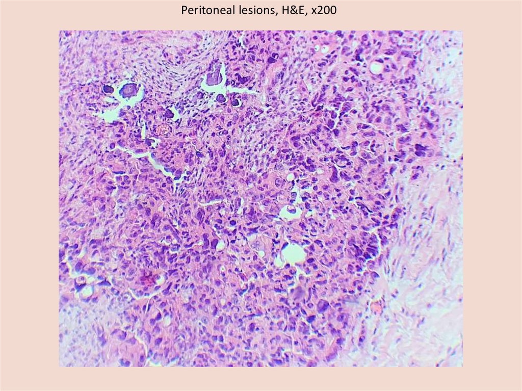
Peritoneum• Visceral and parietal sheets of the peritoneum are dull, moist
• On the surface there are multiple grey thread-like overlays.
• Multiple nodules , gray-black on section, which have solid
consistency with a diameter of 0.3 to 1.5 cm in the parietal and
visceral sheets, merging in places into conglomerates up to 4 cm
8.
Lungs• In the right pleural cavity - 200 ml, in the left - 700 ml of a
clear yellowish liquid
•The mucousa of the trachea and bronchi is pale pink
•The airiness is increased in the upper lobes, with pressure,
folds that do not expand for a long time remain
•Pleura: thin, smooth, shiny, translucent
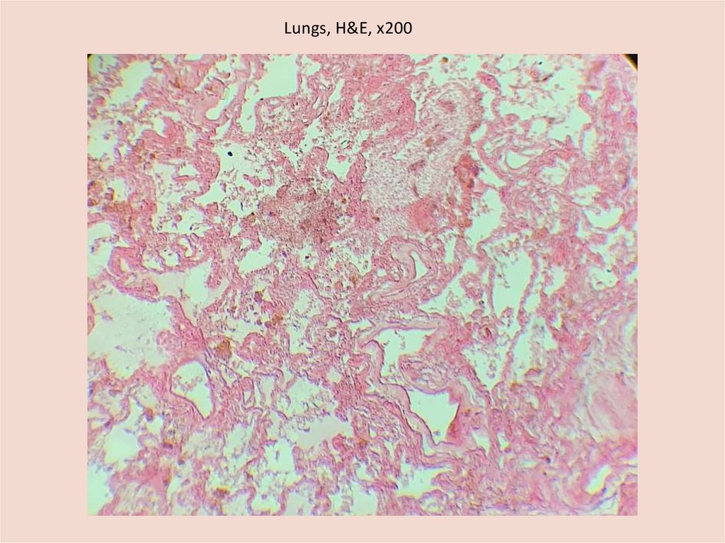
•The lung tissue on the section is red, a large amount of
foaming pinkish fluid flows from the cut, there are extensive
hemorrhages in the lower lobes of the right and left lungs,
mainly subpreleurally.
9.
Lungs•Bronchi: protrude
above the cut
surface, their walls
are thickened,
dense, whitish
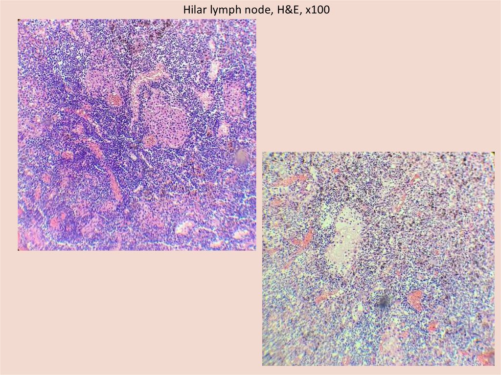
•Hilar and
paratracheal lymph
nodes are gray-black
on the cu, the
maximum size of is
2x1x1 cm.
10.
Large vessels•Aorta: intima of the
thoracic and abdominal
region with yellow
plaques, fibrous
plaques, up to 0.5 cm .
•Pulmonary arteries:
intima is smooth, ivory
colour.
• Large veins: liquid
blood in the lumen
11.
Gastrointestinal tract•Esophagus: mucousa is gray, with longitudinal foldings
•Veins of the lower third are not dilated
•Stomach: mucousa is pale gray, with foldedings, in the
lumen there is a small amount of digested food
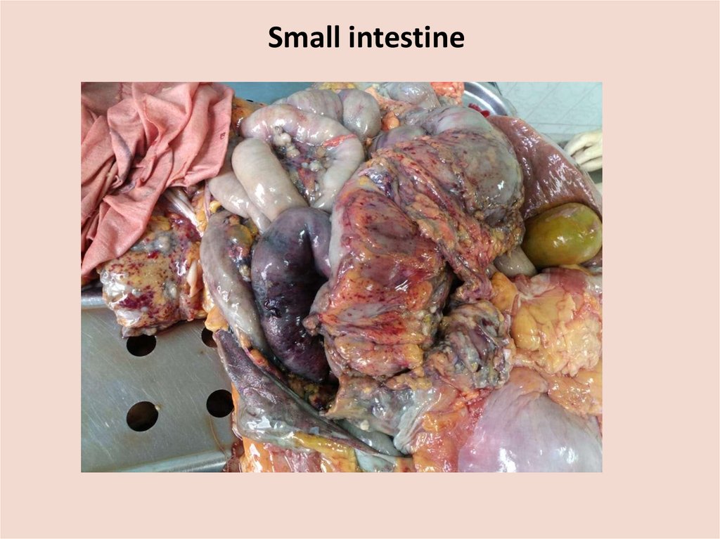
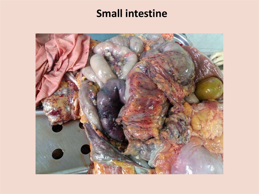
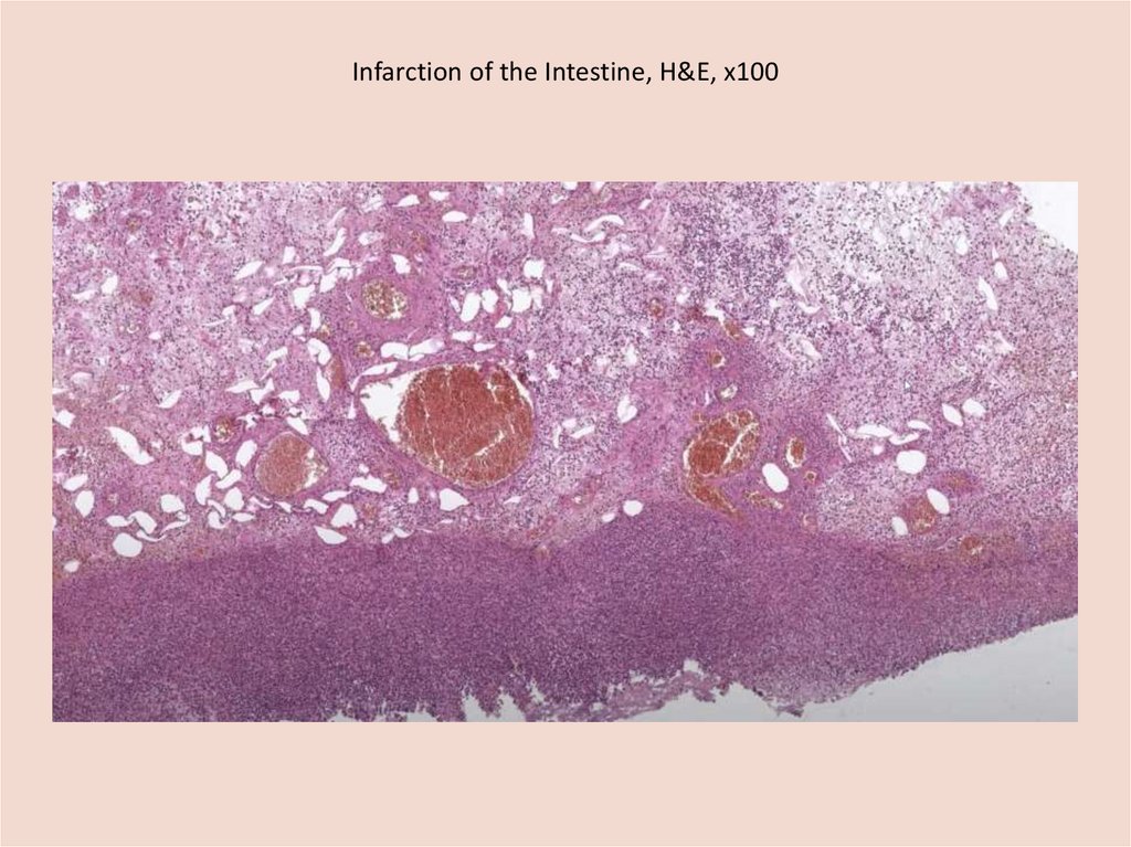
• Small intestine:the loop of the small intestine in the
upper third of the purple color, the intestinal wall,
edematous, flabby consistency, the zone of
demarcation inflammation is not clearly expressed.
Section revialed a thrombus in the lumen of the
branch of the superior mesenteric artery. rest of the
mucous membrane is gray, smoothed, in the lumen
there are liquid yellowish-brown masses.
•Сolon: the mucous membrane is pale gray, folded, the
lumen is filled with faeces.
12.
Small intestine13.
Small intestine14.
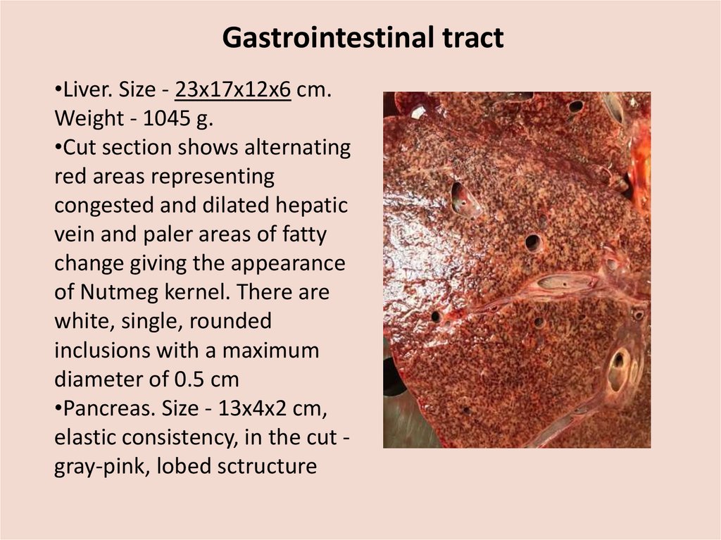
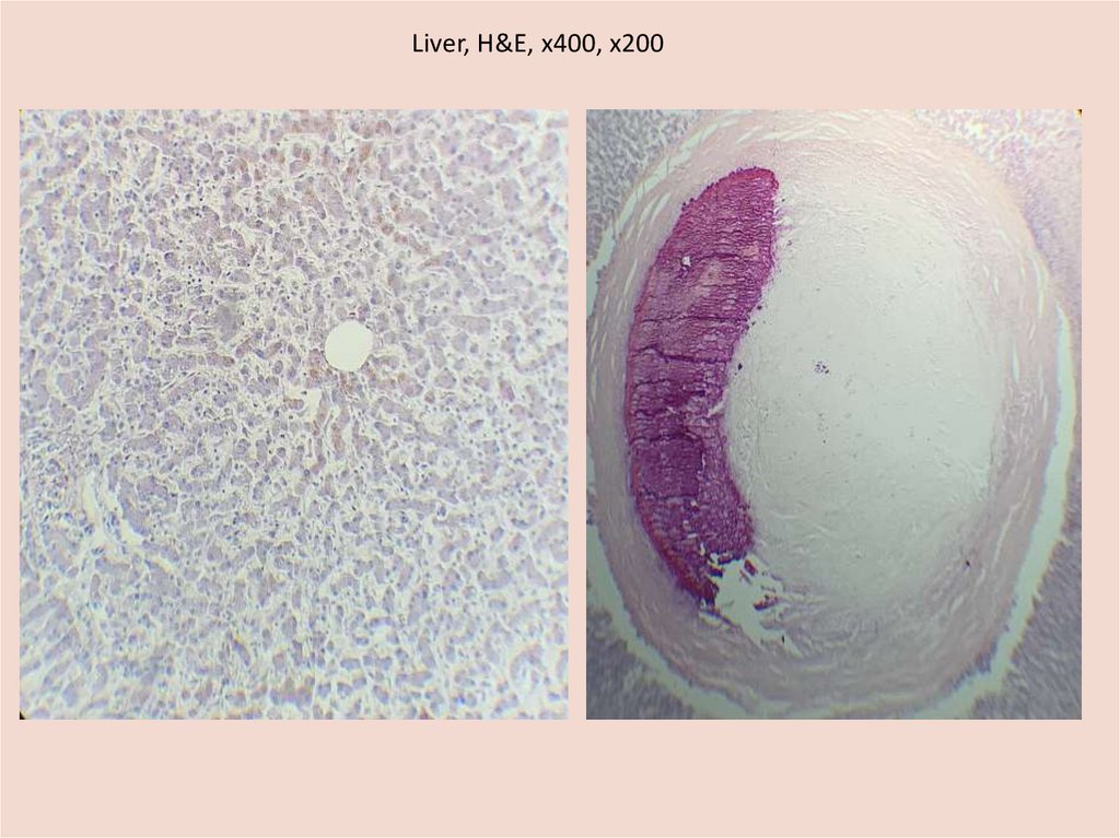
Gastrointestinal tract•Liver. Size - 23х17х12х6 cm.
Weight - 1045 g.
•Cut section shows alternating
red areas representing
congested and dilated hepatic
vein and paler areas of fatty
change giving the appearance
of Nutmeg kernel. There are
white, single, rounded
inclusions with a maximum
diameter of 0.5 cm
•Pancreas. Size - 13x4x2 cm,
elastic consistency, in the cut gray-pink, lobed sctructure
15.
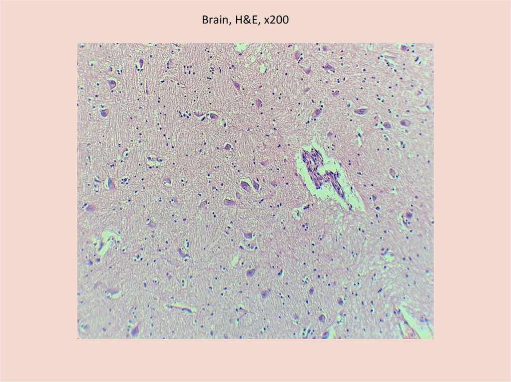
Brain• Size – 17х16х7 cm, weight – 1067 g.
•Consistence: soft, flabby
•The major vessels at the base of the brain have a
usual anatomic distribution and there is no
atherosclerosis.
• The border between gray and white matter is
preserved
•The ventricles are not dilated, contain a transparent
cerebrospinal fluid.
16.
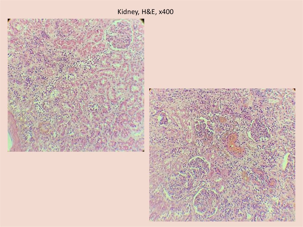
Kidneys•Size: left kidney - 10,5х5х3 cm, right
kidney – 9,5x4x3 cm
•Weight: left kidney - 100 g, right - 140 g
•On the cross sectioning have dense
consistency, a smooth surface
17.
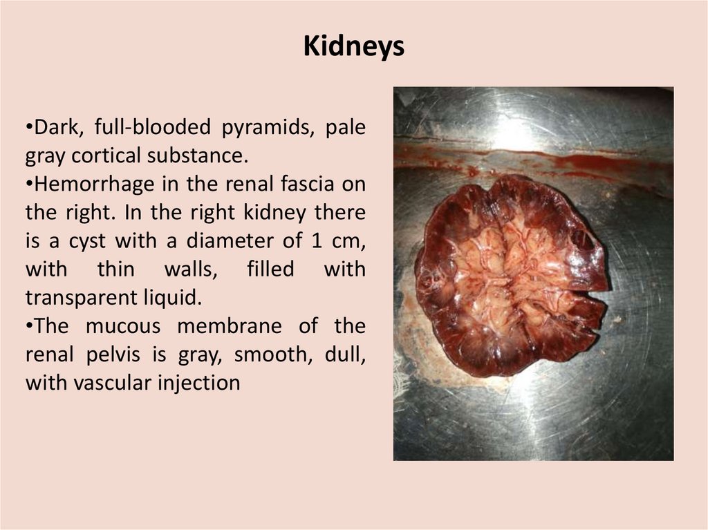
Kidneys•Dark, full-blooded pyramids, pale
gray cortical substance.
•Hemorrhage in the renal fascia on
the right. In the right kidney there
is a cyst with a diameter of 1 cm,
with thin walls, filled with
transparent liquid.
•The mucous membrane of the
renal pelvis is gray, smooth, dull,
with vascular injection
18.
Organs of the urogenital system• Bladder. The mucous membrane is grey, has
foldings.
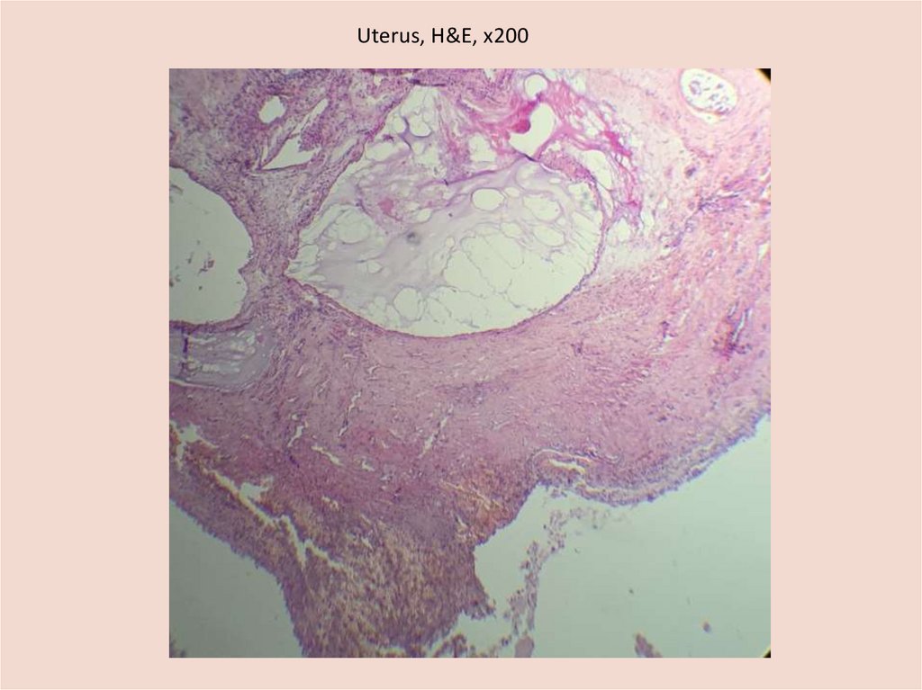
•Uterus - 7x3x2cm, grey, has firm consistency,
with is a cyst d=0.7 cm in the mucous
membrane .
•And a neoplasm, which has grey colour and a
thick pedicle, size - 1x0.5x0.5 cm
• Fallopian tubes and ovaries : are resected
19.
Splin• Size - 11x8x3 cm
• Weight - 112 gWith a smooth capsule
• Flabby consistency
•Dark cherry colour on section
• The character of pulp scraping is
insignificant
20.
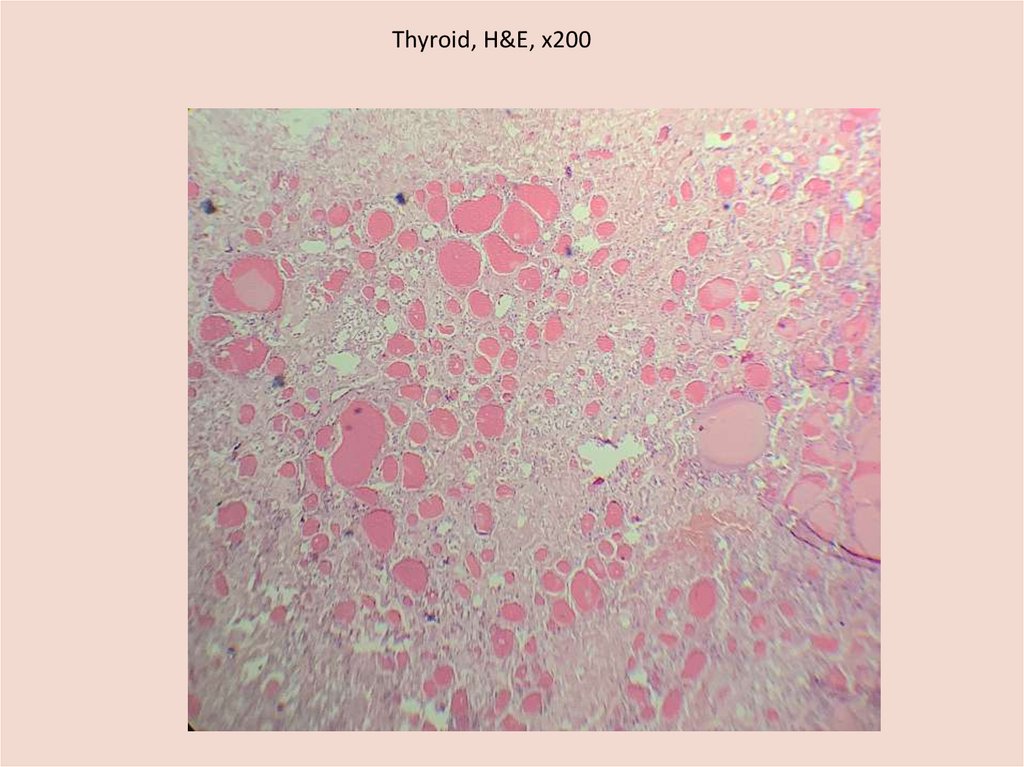
Endocrine system•Thyroid. Size: the right lobe – 1,5x1x1 cm, the
left lobe – is resected, elastic consistency, the
surface is smooth, brown, on the cut - finegrained, brown.
• Adrenal glands. Leaf-shaped, the cortex is
yellow, the medulla is brown.
21.
Microscopic examination22.
Peritoneal lesions, H&E, х10023.
Peritoneal lesions, H&E, х20024.
Peritoneal lesions, H&E , х0025.
Peritoneal lesions, H&E , х40026.
Mesenteric lymph node, H&E , х40027.
Heart, H&E, х20028.
Heart, H&E, х40029.
Heart, H&E, х20030.
Infarction of the Intestine, H&E, х10031.
Lungs, H&E, х20032.
Lungs, H&E, х20033.
Lungs, H&E, х20034.
Lungs, H&E, х20035.
Hilar lymph node, H&E, х10036.
Brain, H&E, х20037.
Liver, H&E, х400, x20038.
Kidney, H&E, х40039.
Uterus, H&E, х20040.
Thyroid, H&E, х20041.
Final clinical diagnosis:The underlying disease. Ovarian cancer T3N1M1, III A2: histologically - undifferentiated
endometrioid adenocarcinoma RE4 +, HER 2-neu-negative. Carcinomatosis of the
peritoneum. Ascites. Metastasis in S 10 of the right lung. 2 courses of PCT according to the
RS scheme from July 2020 to September 2020.
Concomitant disease. Ischemic heart disease: angina pectoris II FC. Postinfarction
cardiosclerosis from 2012 and 2018. RCA stenting in the distal region, aspiration of thrombi
from the PTA RCA in the distal region from November 17, 2018
Background disease. Hypertension stage III, grade 3, risk of CVC 4.
Accompanying illnesses. D12.5 Sigmoid tubulovillous adenoma with moderate epithelial
dysplasia.
Accompanying illnesses. Chronic pyelonephritis. Nodular goiter. Hypothyroidism
Hemithyroidectomy on the left, resection of the right lobe of the vanilla from 2008.
Arthrosis of both shoulder joints.
Complications. Acute pulmonary heart failure. Tumor intoxication. Mild anemia.
Myocardial infarction type II. Thromboembolism of the branches of the pulmonary artery.
Stress-induced erosion and stomach ulcers with possible development of gastrointestinal
bleeding. Bilateral hydrothorax. Pulmonary edema. Cerebral edema. Resuscitation
measures. Indirect cardiac massage
42.
Pathological diagnosis (primary):The underlying disease. Ovarian cancer with parietal and visceral peritoneum
carcinomatosis, liver metastases Operation of laparoscopic bilateral adnexectomy
(date unknown). 2 courses of polychemotherapy from 08.2020. pTxN1M1.
Complications. Thrombosis of the branch of the superior mesenteric artery. Wet
gangrene of the loop of the small intestine. Fibrinous peritonitis. Shock of mixed
etiology. Hemorrhagic syndrome: multiple petechial hemorrhages on the skin,
parietal and visceral peritoneum; imbibition with blood of the lower lobes of both
lungs. Fatty degeneration of the liver and myocardium. Shock kidneys. Ascites
(3000 ml). Bilateral hydrothorax (700 ml in the left pleural cavity, 200 ml in the
right pleural cavity). Pulmonary edema. Cerebral edema.
Resuscitation measures: chest compressions.
43.
Pathological diagnosis (primary):Accompanying illnesses. Chronic obstructive mucous bronchitis. Diffuse reticular
pneumosclerosis. Chronic obstructive pulmonary emphysema. Large focal cardiosclerosis
in the lateral wall of the left ventricle. Atherosclerosis of the aorta (fat spots and stripes),
coronary arteries of the heart (fat spots and stripes). Operations of stenting of the right
coronary artery in the distal part, aspiration of blood clots from the posterior lateral
branch of the right coronary artery, stenting of the posterior lateral branch of the right
coronary artery in the distal part, 11/17/2018 (clinically). Nodular goiter (clinically).
Operation of hemithyroidectomy on the left, resection of the right lobe of the thyroid
gland (2008). Endometrial polyp.
The clinical and pathological diagnoses coincided. Notes on patient management: the last
blood test performed was dated 08/28/2020, the date of death was 09/15/2020.
The immediate cause of death is peritonitis.
44.
CLINICOPATHOLOGIC CORRELATIONPatient N, 67 years old, suffered from ovarian cancer with carcinomatosis of the
peritoneum and metastases to the liver for a long time, the patient underwent
bilateral laparoscopic adnexectomy (at autopsy: both fallopian tubes and both
ovaries were absent, on the anterior abdominal wall there were scars from
laparoscopic access). The patient developed tumor coagulopathy, manifested by
thrombosis of the coronary arteries of the heart in the distal regions, which led to
the development of myocardial infarction and later - large-focal cardiosclerosis, as
well as thrombosis of the superior mesenteric artery branch, complicated by
moist gangrene of the loop of the small intestine and fibrinosis. The immediate
cause of death is fibrinous peritonitis. The peculiarity of the case is a pronounced
tumor coagulopathy, which led to the development of large-focal cardiosclerosis
and intestinal gangrene.
45.
.Pathological diagnosis after histological
examination:
The histological examination confirmed the pathological
diagnosis. Clarified the nature of the focus in the liver - a
focus of sclerosis with calcification.