


 Медицина
Похожие презентации:
")


Dermatology
1.
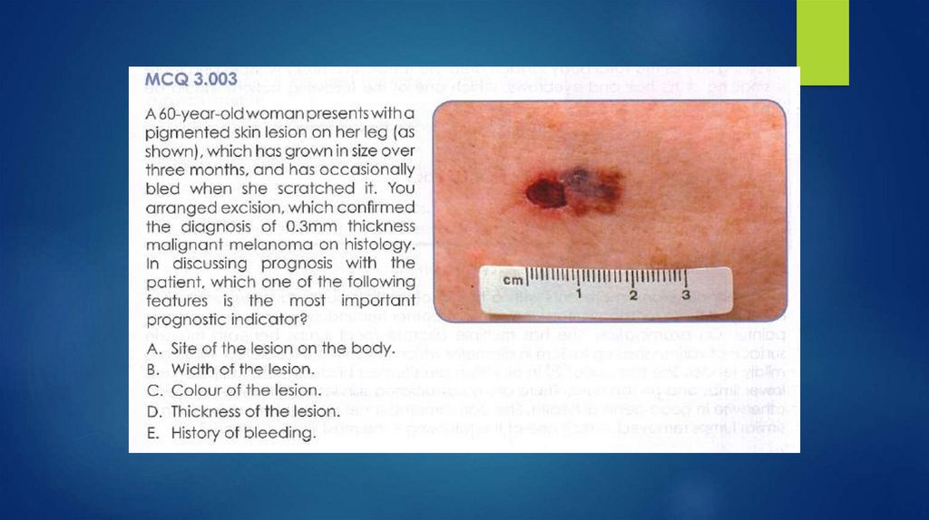
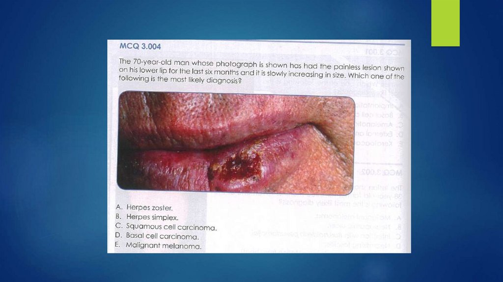
DermatologyHANDBOOK 3.003 – 3.009
2.
3.
4.
5.
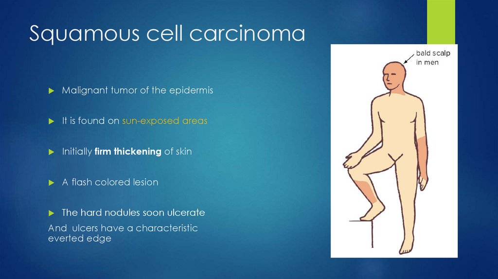
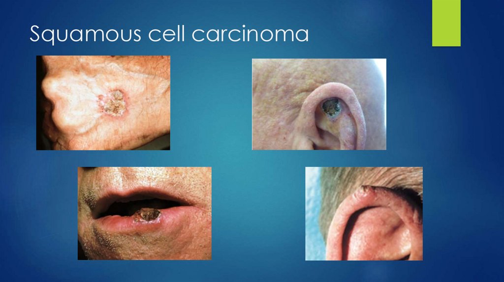
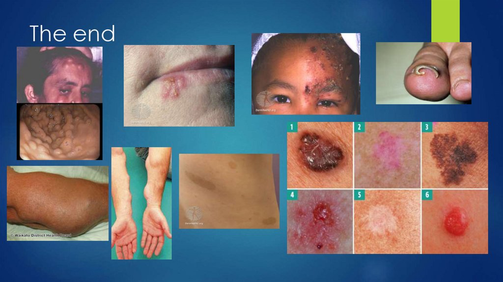
Squamous cell carcinomaMalignant tumor of the epidermis
It is found on sun-exposed areas
Initially firm thickening of skin
A flash colored lesion
The hard nodules soon ulcerate
And ulcers have a characteristic
everted edge
6.
Squamous cell carcinoma7.
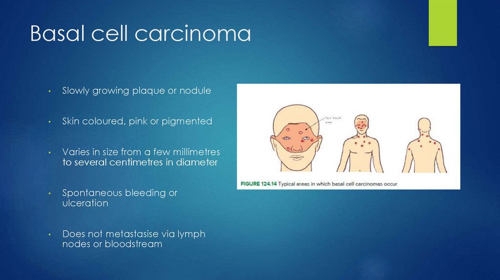
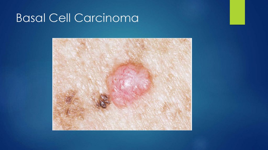
Basal cell carcinomaSlowly growing plaque or nodule
Skin coloured, pink or pigmented
Varies in size from a few millimetres
to several centimetres in diameter
Spontaneous bleeding or
ulceration
Does not metastasise via lymph
nodes or bloodstream
8.
Basal Cell Carcinoma9.
Basal Cell Carcinoma10.
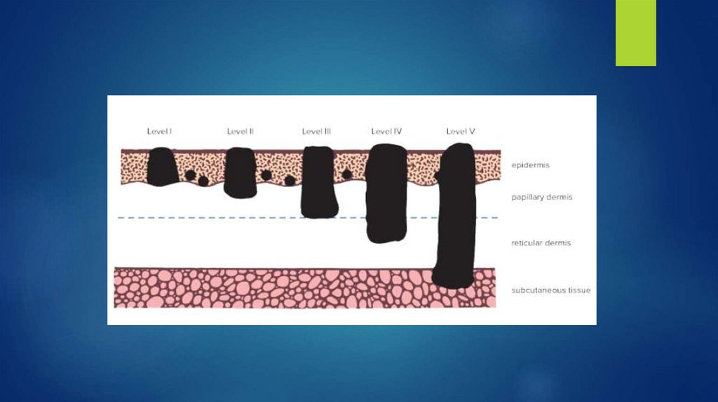
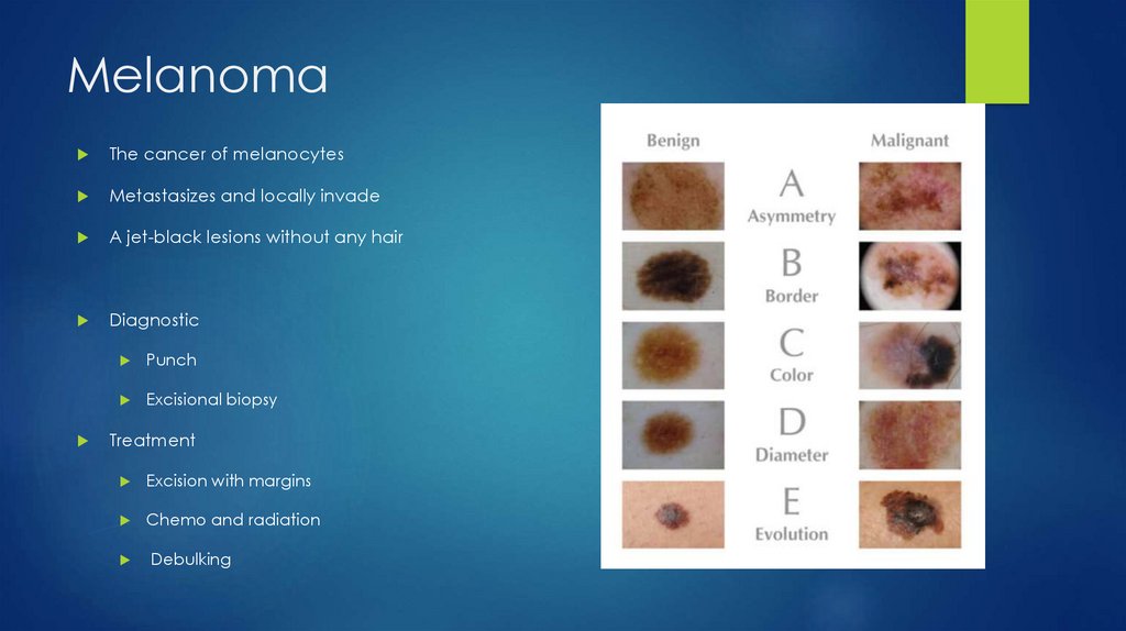
MelanomaThe cancer of melanocytes
Metastasizes and locally invade
A jet-black lesions without any hair
Diagnostic
Punch
Excisional biopsy
Treatment
Excision with margins
Chemo and radiation
Debulking
11.
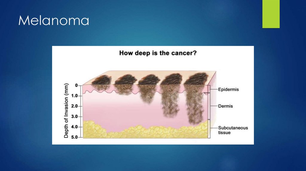
Melanoma12.
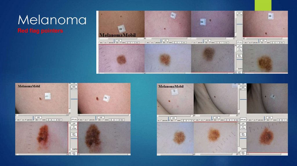
MelanomaRed flag pointers
13.
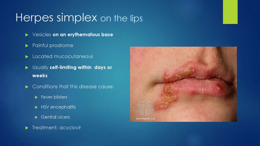
Herpes simplex on the lipsVesicles on an erythematous base
Painful prodrome
Located mucocutaneous
Usually self-limiting within days or
weeks
Conditions that this disease cause:
Fever blisters
HSV encephalitis
Genital ulcers
Treatment: acyclovir
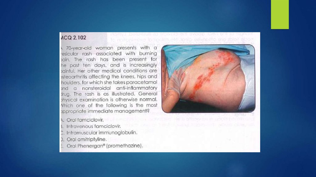
14.
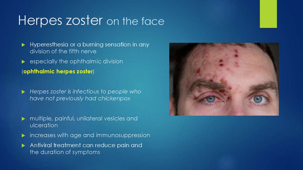
Herpes zoster on the faceHyperesthesia or a burning sensation in any
division of the fifth nerve
especially the ophthalmic division
(ophthalmic herpes zoster)
Herpes zoster is infectious to people who
have not previously had chickenpox
multiple, painful, unilateral vesicles and
ulceration
increases with age and immunosuppression
Antiviral treatment can reduce pain and
the duration of symptoms
15.
16.
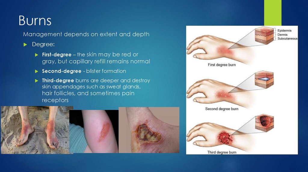
BurnsManagement depends on extent and depth
Degree:
First-degree – the skin may be red or
gray, but capillary refill remains normal
Second-degree - blister formation
Third-degree burns are deeper and destroy
skin appendages such as sweat glands,
hair follicles, and sometimes pain
receptors
17.
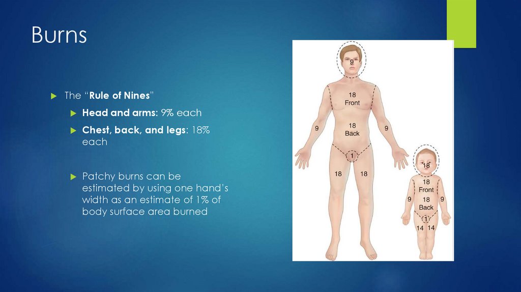
BurnsThe “Rule of Nines”
Head and arms: 9% each
Chest, back, and legs: 18%
each
Patchy burns can be
estimated by using one hand’s
width as an estimate of 1% of
body surface area burned
18.
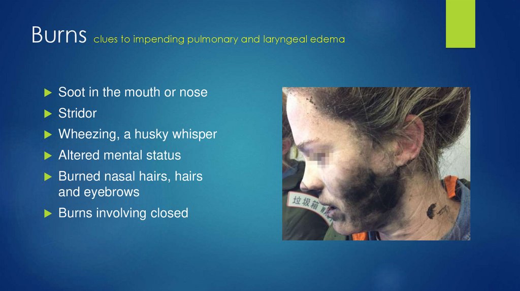
Burns clues to impending pulmonary and laryngeal edemaSoot in the mouth or nose
Stridor
Wheezing, a husky whisper
Altered mental status
Burned nasal hairs, hairs
and eyebrows
Burns involving closed
19.
Burn treatmentIf patient has signs of severe respiratory injury, the first step is to intubate before
more severe laryngeal edema can occur and make the intubation difficult.
If carboxyhemoglobin level is significantly elevated (>5–10%), administer 100%
oxygen.
Fluid resuscitation over the first 24 hours.
Use Ringer’s lactate as the preferred fluid
Afterward, when the diffuse capillary leak improves, give enough fluid to
maintain urine output >0.5–1 mL per kg per hour.
Give stress ulcer prophylaxis with H2 blocker or PPI.
To prevent infection, use topical treatment with silver sulfadiazine.
Do not break blisters and do not use steroids.
20.
21.
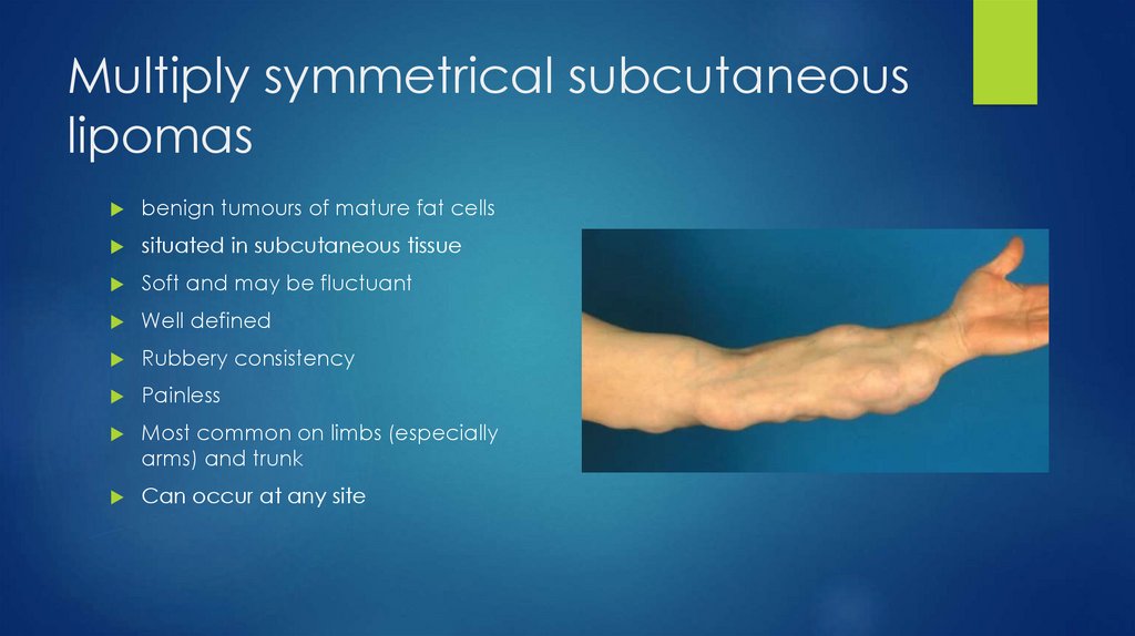
Multiply symmetrical subcutaneouslipomas
benign tumours of mature fat cells
situated in subcutaneous tissue
Soft and may be fluctuant
Well defined
Rubbery consistency
Painless
Most common on limbs (especially
arms) and trunk
Can occur at any site
22.
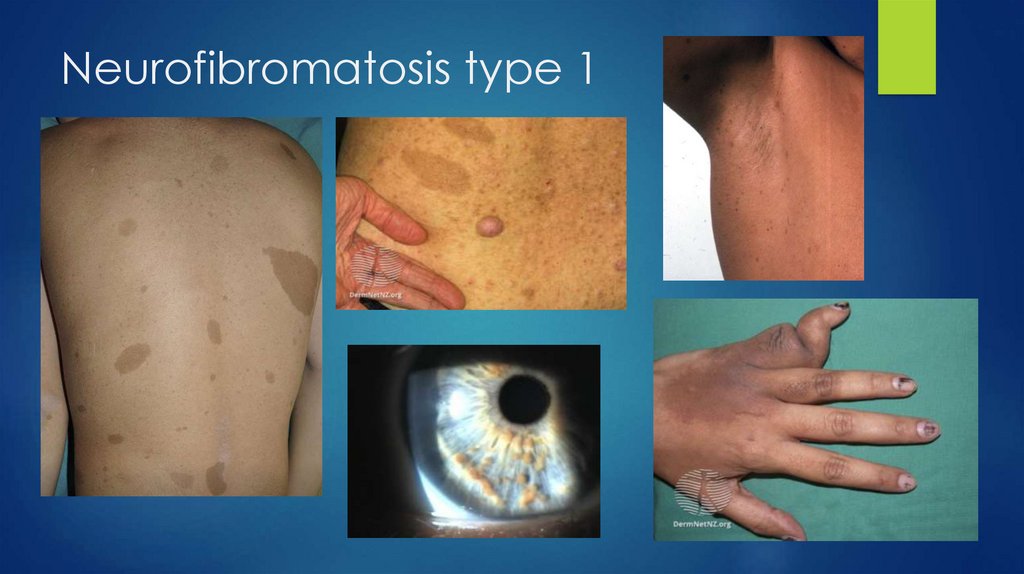
Neurofibromatosis type 1von Recklinghausen disorder
Clinical features
Six or more café-au-lait spots
Freckling in the axillary or inguinal regions
Flesh-coloured cutaneous tumours
Hypertension
Iris hamartomas
Learning difficulty
Musculoskeletal problems
Optic nerve gliomas
23.
Neurofibromatosis type 124.
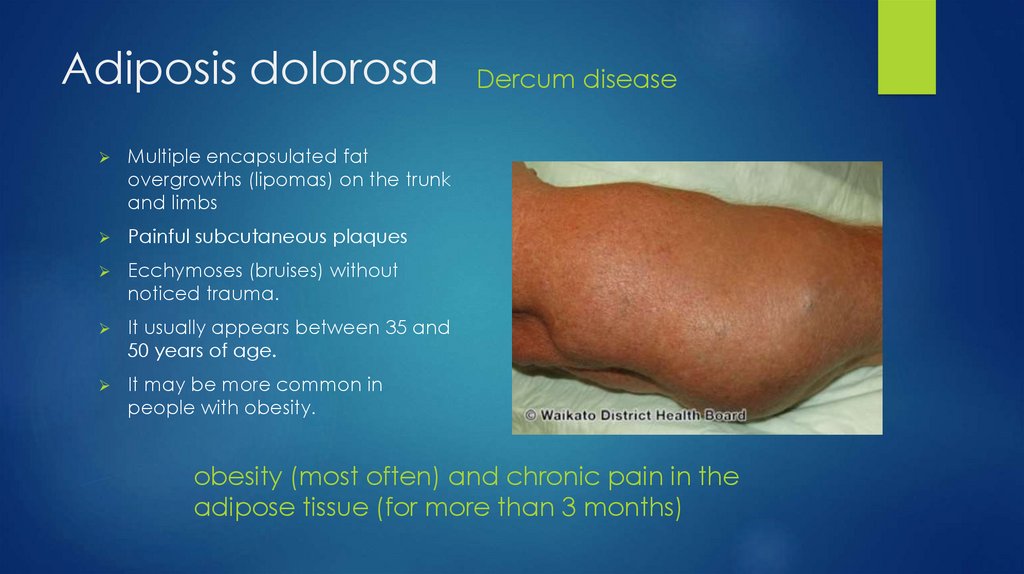
Adiposis dolorosaMultiple encapsulated fat
overgrowths (lipomas) on the trunk
and limbs
Painful subcutaneous plaques
Ecchymoses (bruises) without
noticed trauma.
It usually appears between 35 and
50 years of age.
It may be more common in
people with obesity.
Dercum disease
obesity (most often) and chronic pain in the
adipose tissue (for more than 3 months)
25.
Dermoid cystThe most common location for
dermoid cysts is the lateral third of
the eyebrows; however, they also
may occur on the mid forehead,
scalp, nose, anterior neck, and
trunk.
they are caused by the
implantation of epithelial tissue
into another structure
dermoid cysts are made up of
epidermal and dermal
components: keratinocytes, hair
follicles and hair, and sweat
glands
Epidermoid cyst
Are similar in structure and origin to
dermoid tumors and the two are
often grouped together.
Epidermoid tumors are lined with
stratified squamous epithelium
(skin) as dermoids are, but do not
contain the additional skin
appendages
26.
27.
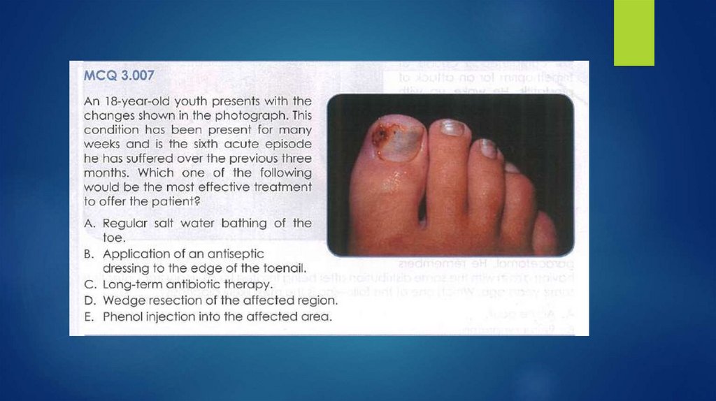
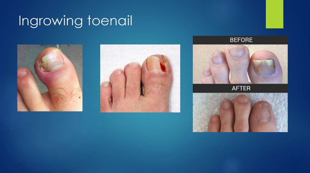
Ingrowing toenailthe sides or corner of the toenail digs into the skin at the end or side
of the toe
Mostly affects the outer edge of the big toe
Causes: ill-fitting shoes, improper trimming of toenails, injury near the
nail, fungal infections of the nail, prescribed medications, abnormal
nail shape
28.
Ingrowing toenail29.
The end30.
31.
32.
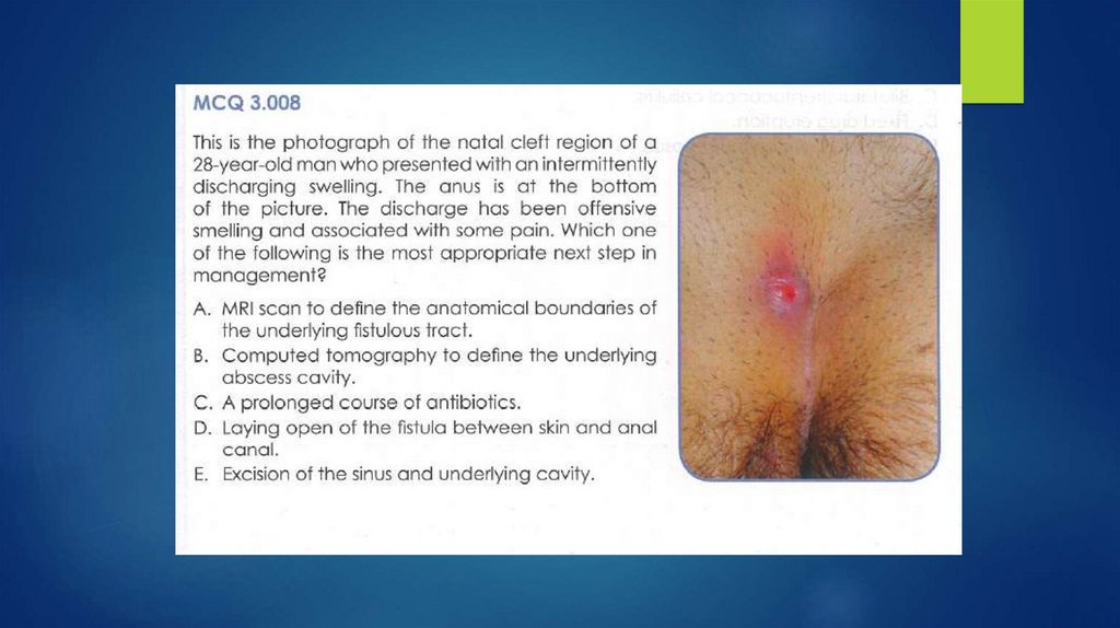
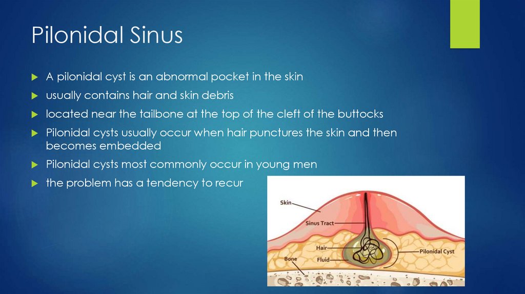
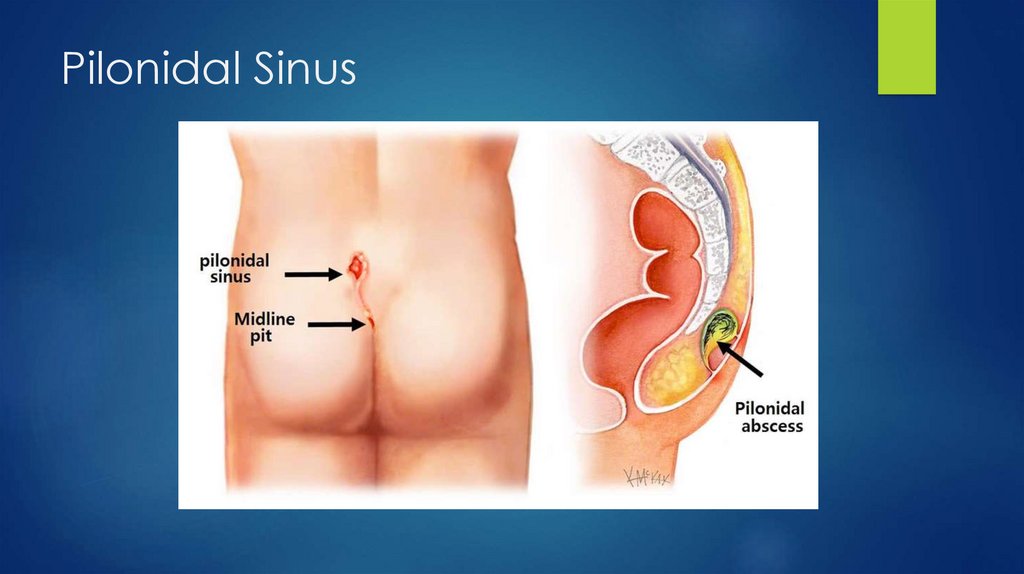
Pilonidal SinusA pilonidal cyst is an abnormal pocket in the skin
usually contains hair and skin debris
located near the tailbone at the top of the cleft of the buttocks
Pilonidal cysts usually occur when hair punctures the skin and then
becomes embedded
Pilonidal cysts most commonly occur in young men
the problem has a tendency to recur
33.
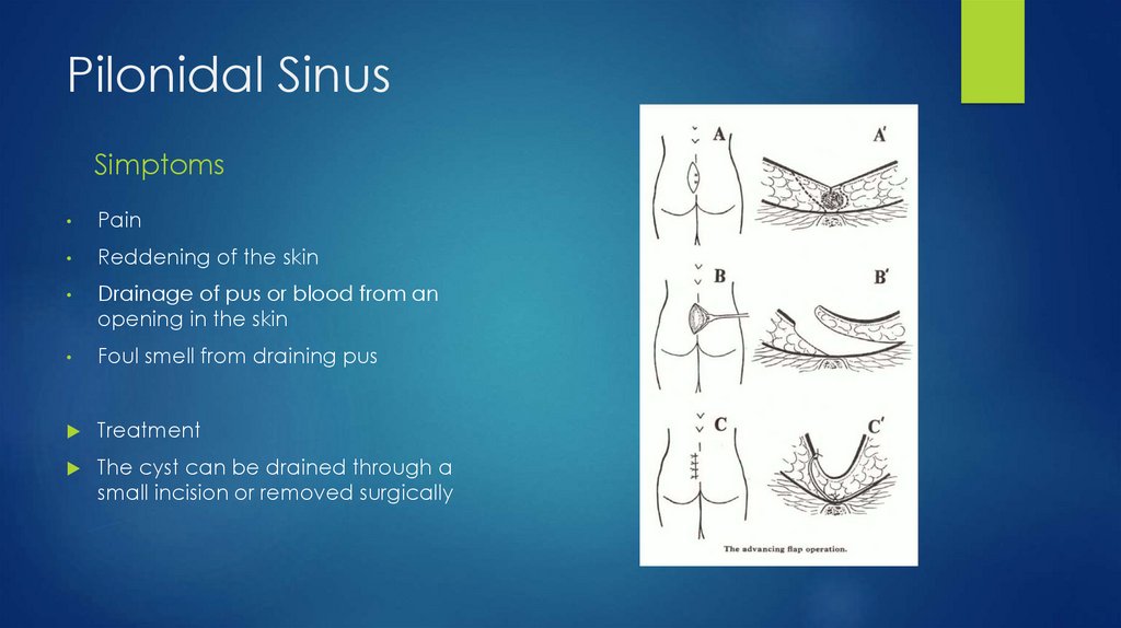
Pilonidal SinusSimptoms
Pain
Reddening of the skin
Drainage of pus or blood from an
opening in the skin
Foul smell from draining pus
Treatment
The cyst can be drained through a
small incision or removed surgically
34.
Pilonidal Sinus35.
36.
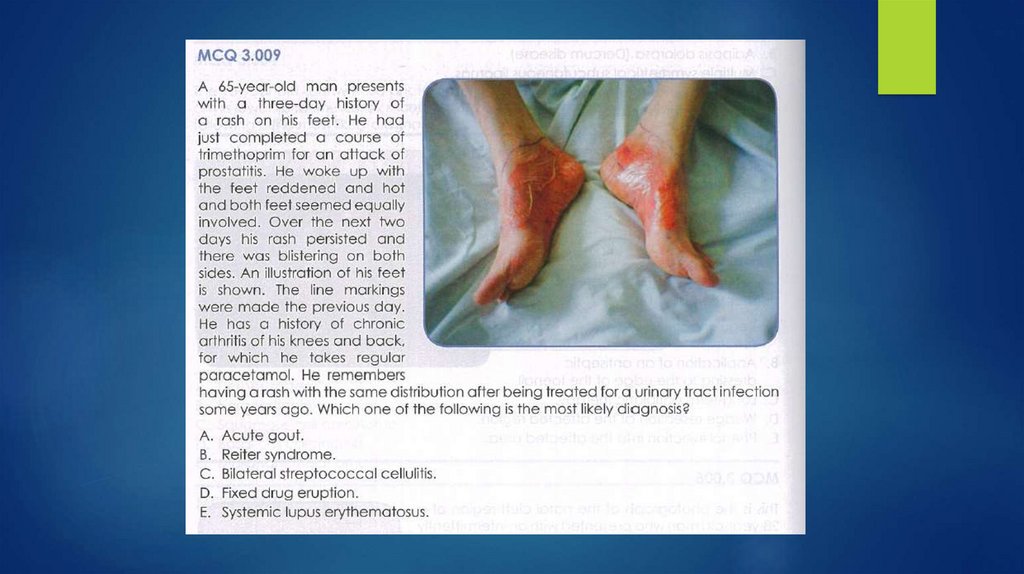
Fixed drug eruptiontrimethoprim
Drugs with the highest skin reaction
rates
The mechanism of fixed drug
eruption is unknown
Penicillin and derivatives
Sulphonamides*
The most commonly affected areas
are the face, hands and genitalia
Trimethoprim*
appearance within hours of the
drug’s administration
Thiazide diuretics
Allopurinol*
Dapsone*
NSAIDs, esp. piroxicam*
Treatment
To recognise the offending agent
Nevirapine*, abacavir*
and withdraw it
Barbiturates
Quinidine
Anti-epileptics (phenytoin, lamotrigine*)
Blood products
Gold salts
The rash should be treated
according to its nature
37.
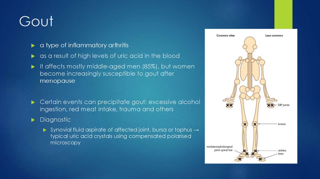
Gouta type of inflammatory arthritis
as a result of high levels of uric acid in the blood
It affects mostly middle-aged men (85%), but women
become increasingly susceptible to gout after
menopause
Certain events can precipitate gout: excessive alcohol
ingestion, red meat intake, trauma and others
Diagnostic
Synovial fluid aspirate of affected joint, bursa or tophus →
typical uric acid crystals using compensated polarised
microscopy
38.
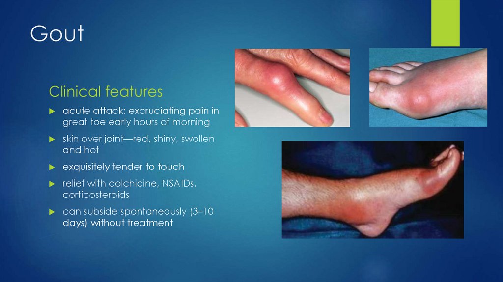
GoutClinical features
acute attack: excruciating pain in
great toe early hours of morning
skin over joint—red, shiny, swollen
and hot
exquisitely tender to touch
relief with colchicine, NSAIDs,
corticosteroids
can subside spontaneously (3–10
days) without treatment
39.
Goutgood advice and patient education information
provision of rapid pain relief
preventing further attacks
prevention of destructive arthritis and tophi dealing with precipitating
factors and comorbid conditions
The acute attack
NSAIDs (except aspirin), in full dosage
Corticosteroids: prednisolone
Colchicine: colchicine
Prevention: Allopurinol
40.
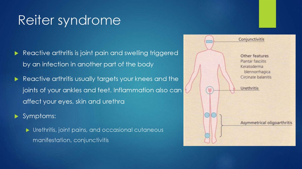
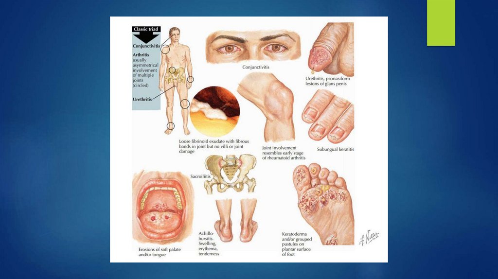
Reiter syndromeReactive arthritis is joint pain and swelling triggered
by an infection in another part of the body
Reactive arthritis usually targets your knees and the
joints of your ankles and feet. Inflammation also can
affect your eyes, skin and urethra
Symptoms:
Urethritis, joint pains, and occasional cutaneous
manifestation, conjunctivitis
41.
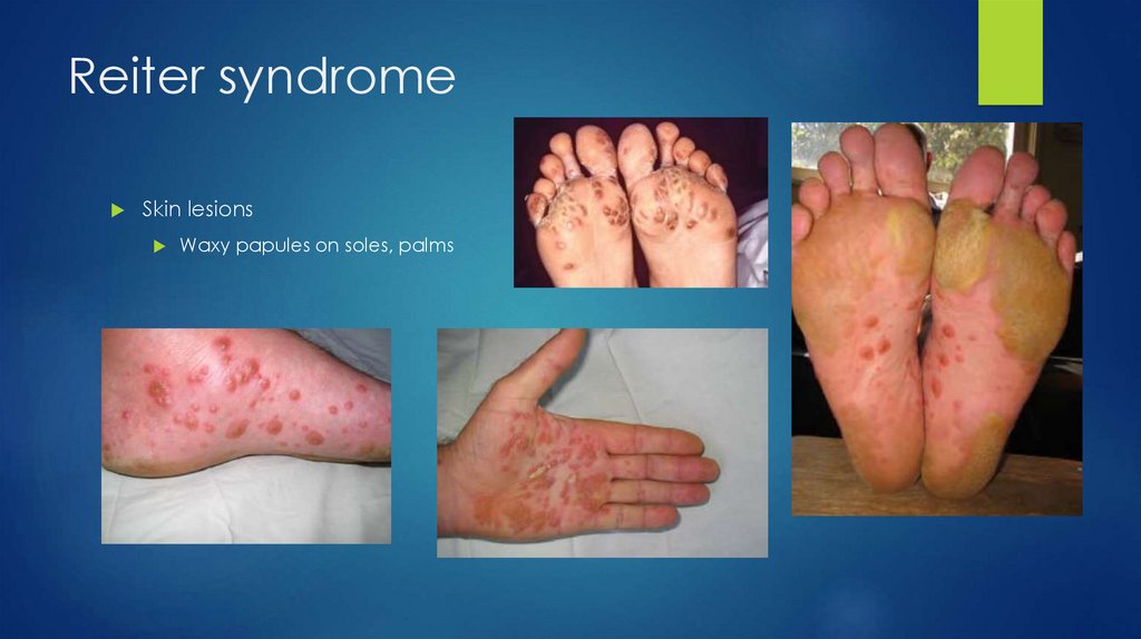
Reiter syndromeSkin lesions
Waxy papules on soles, palms
42.
43.
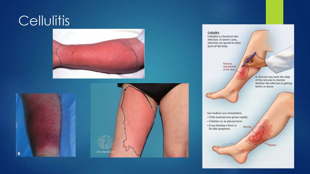
Bilateral streptococcal cellulitisCellulitis is a common bacterial infection
a localised area of red, painful, swollen skin, and systemic symptoms
The most common bacteria causing cellulitis are Streptococcus
pyogenes (two-thirds of cases) and Staphylococcus aureus (one
third)
Clinical features:
Cellulitis can affect any site, most often a limb
It is usually unilateral; a bilateral disease is more often due to another
condition
It can occur by itself or complicate an underlying skin condition or
wound.
44.
Cellulitis45.
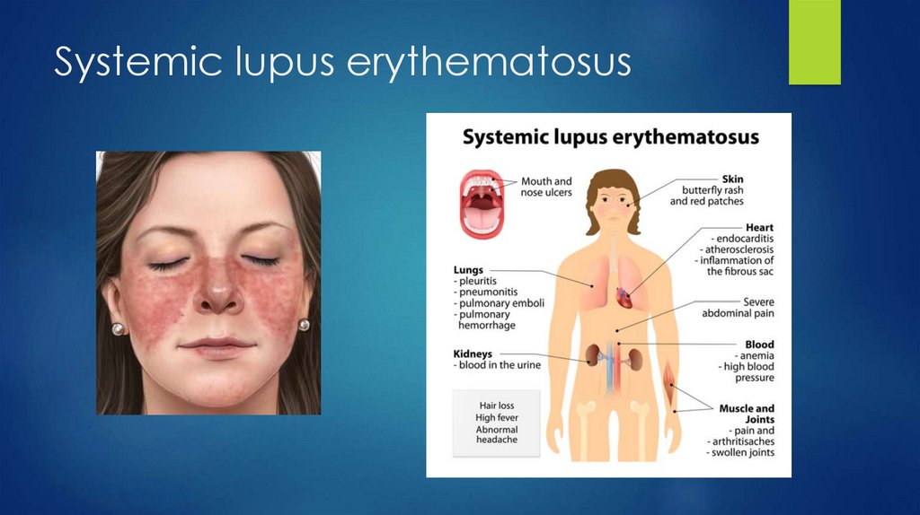
Systemic lupus erythematosus46.
Systemic lupus erythematosusDxT
Polyarthritis + fatigue + skin lesion
47.
48.
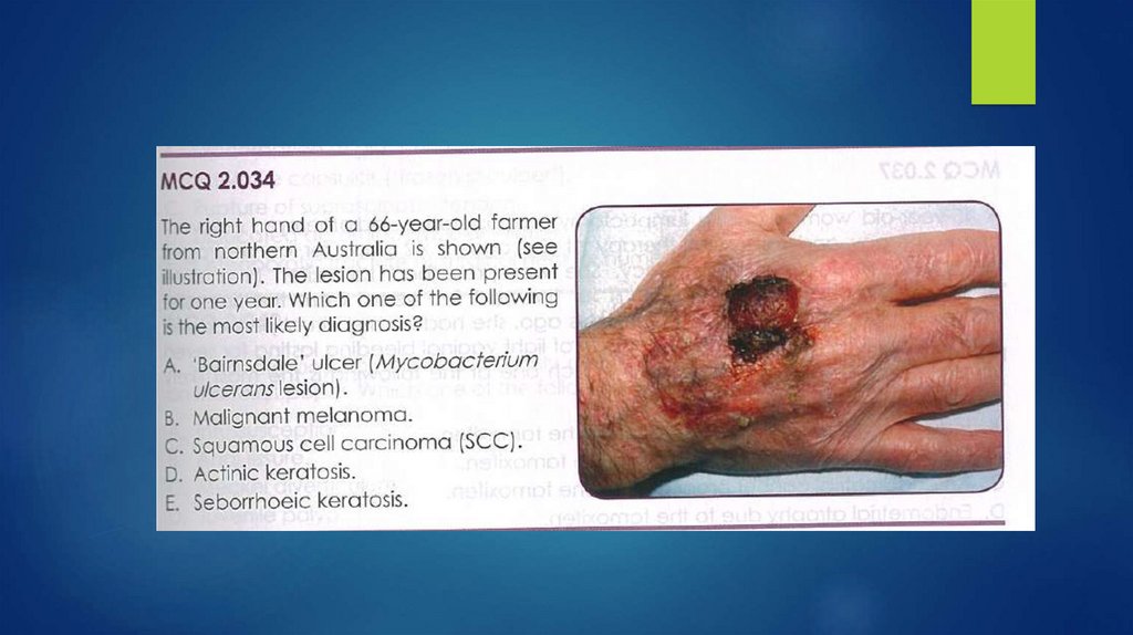
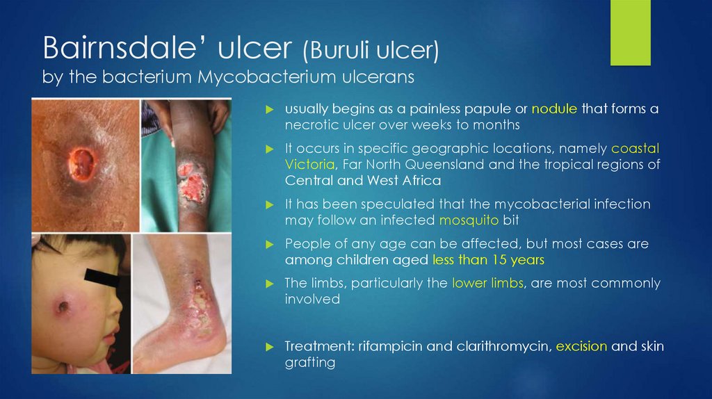
Bairnsdale’ ulcer (Buruli ulcer)by the bacterium Mycobacterium ulcerans
usually begins as a painless papule or nodule that forms a
necrotic ulcer over weeks to months
It occurs in specific geographic locations, namely coastal
Victoria, Far North Queensland and the tropical regions of
Central and West Africa
It has been speculated that the mycobacterial infection
may follow an infected mosquito bit
People of any age can be affected, but most cases are
among children aged less than 15 years
The limbs, particularly the lower limbs, are most commonly
involved
Treatment: rifampicin and clarithromycin, excision and skin
grafting
49.
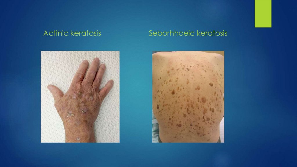
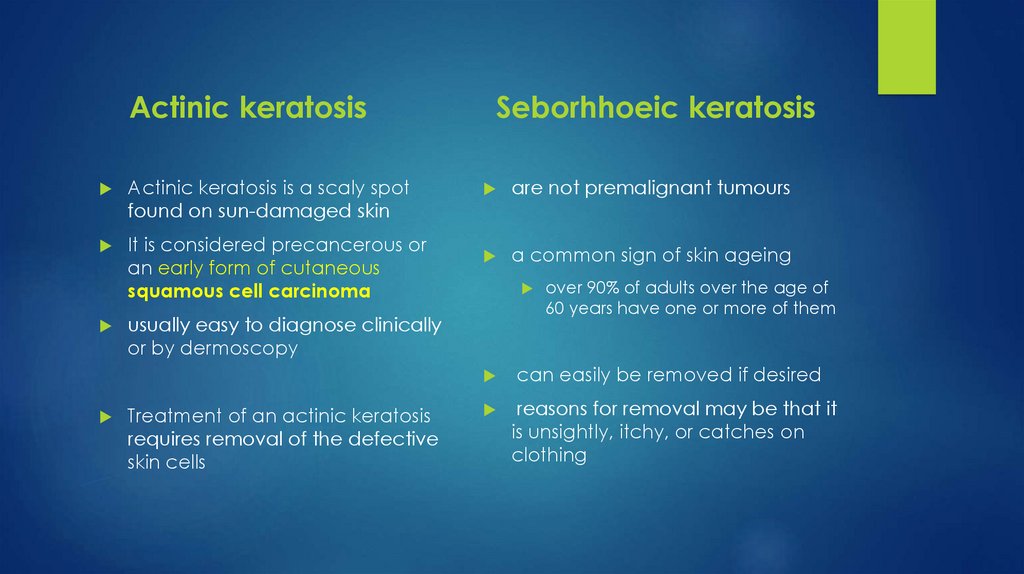
Actinic keratosisSeborhhoeic keratosis
50.
Actinic keratosisActinic keratosis is a scaly spot
found on sun-damaged skin
It is considered precancerous or
an early form of cutaneous
squamous cell carcinoma
Seborhhoeic keratosis
are not premalignant tumours
a common sign of skin ageing
usually easy to diagnose clinically
or by dermoscopy
Treatment of an actinic keratosis
requires removal of the defective
skin cells
over 90% of adults over the age of
60 years have one or more of them
can easily be removed if desired
reasons for removal may be that it
is unsightly, itchy, or catches on
clothing
51.
52.
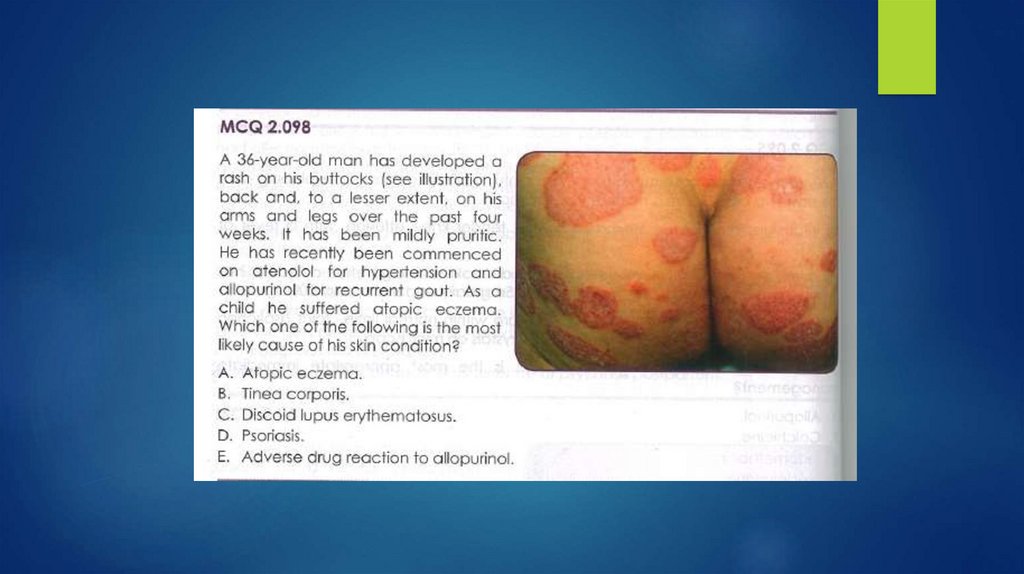
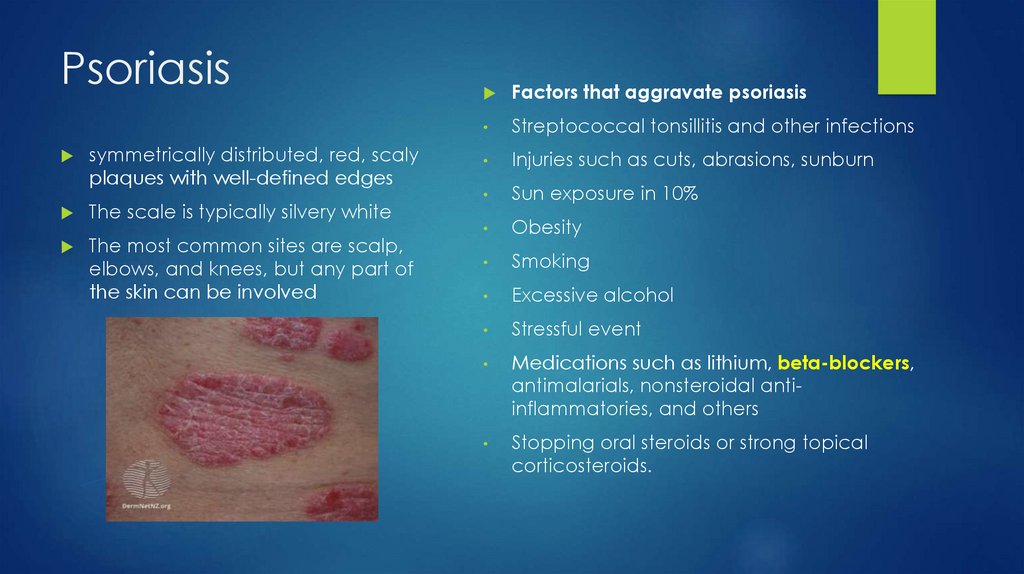
Psoriasissymmetrically distributed, red, scaly
plaques with well-defined edges
The scale is typically silvery white
The most common sites are scalp,
elbows, and knees, but any part of
the skin can be involved
Factors that aggravate psoriasis
Streptococcal tonsillitis and other infections
Injuries such as cuts, abrasions, sunburn
Sun exposure in 10%
Obesity
Smoking
Excessive alcohol
Stressful event
Medications such as lithium, beta-blockers,
antimalarials, nonsteroidal antiinflammatories, and others
Stopping oral steroids or strong topical
corticosteroids.
53.
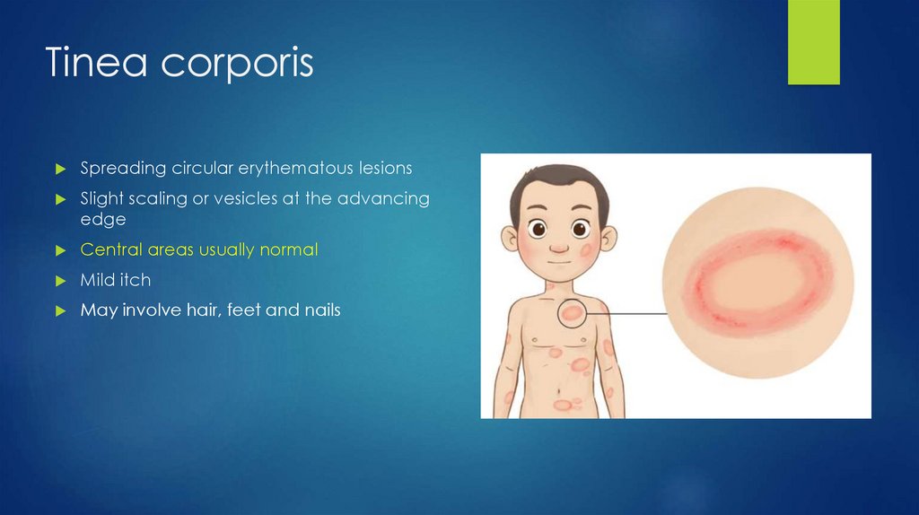
Tinea corporisSpreading circular erythematous lesions
Slight scaling or vesicles at the advancing
edge
Central areas usually normal
Mild itch
May involve hair, feet and nails
54.
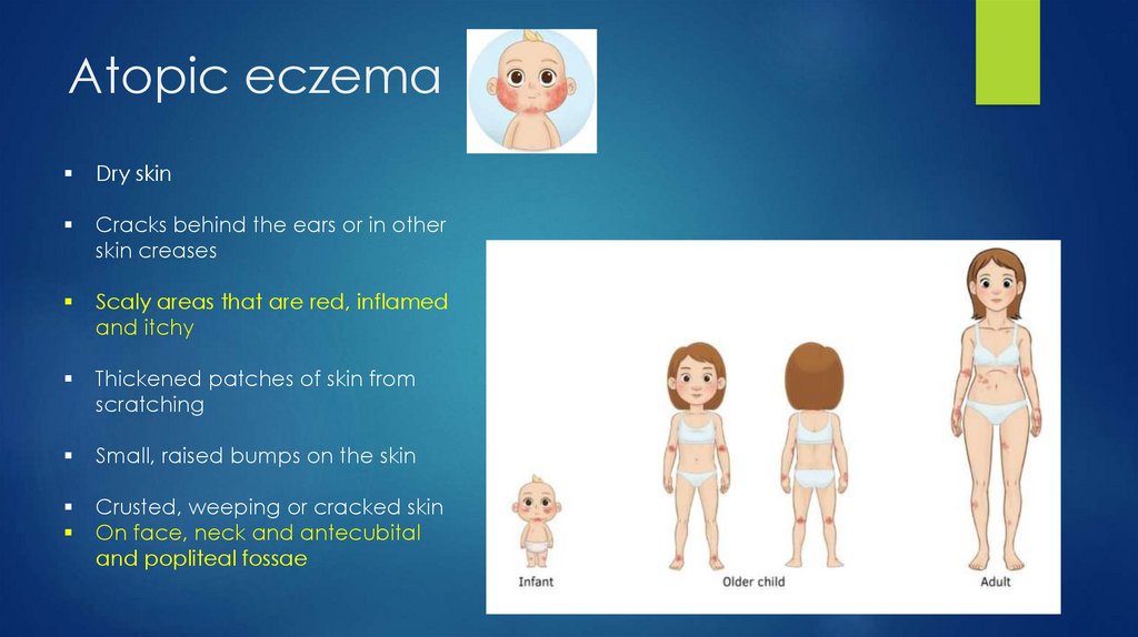
Atopic eczemaDry skin
Cracks behind the ears or in other
skin creases
Scaly areas that are red, inflamed
and itchy
Thickened patches of skin from
scratching
Small, raised bumps on the skin
Crusted, weeping or cracked skin
On face, neck and antecubital
and popliteal fossae
55.
Discoid lupus erithematosusscaly, disk-like plaques on the
scalp, face, and ears that may
cause pigmentary changes,
scarring and hair loss
56.
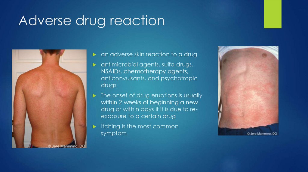
Adverse drug reactionan adverse skin reaction to a drug
antimicrobial agents, sulfa drugs,
NSAIDs, chemotherapy agents,
anticonvulsants, and psychotropic
drugs
The onset of drug eruptions is usually
within 2 weeks of beginning a new
drug or within days if it is due to reexposure to a certain drug
Itching is the most common
symptom