


")


 Медицина
Похожие презентации:

")


Rehabilitation for shoulder fractures & surgeries
1.
بسم هللا الرحمن الرحيم2. REHABILITATION FOR SHOULDER FRACTURES & SURGERIES
REHABILITATION FOR SHOULDERFRACTURES & SURGERIES
• Clavicle fractures
• Proximal head of humerus
fractures
Dr. ahmed Samir Mohamed
Lecturer of physical therapy for orthopedic and its
surgeries
3. Clavicle fractures
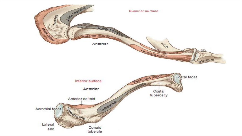
When one looks from above, it is evident that the shaft of the clavicle iscurved, with its anterior surface being generally convex medially and concave
laterally.
Osteologic Features of the Clavicle
• Shaft
• Sternal end
• Costal facet
• Costal tuberosity
• Acromial end
• Acromial facet
• Conoid tubercle
• Trapezoid line
4.
5. Sternoclavicular Joint
The sternoclavicular (SC)joint
is
a
complex
articulation, involving the
medial end of the clavicle, the
clavicular facet on
the
sternum, and the superior
border of the cartilage of the
first rib
6. Arthrokinematics of SCJ
7. Arthrokinematics of SCJ
8. Acromioclavicular joint (ACJ)
Theacromioclavicular
(AC)
joint
is
the
articulation between the
lateral end of the clavicle
and the acromion of the
scapula
9. AC stabilizers
10. Arthrokinematics of AC joint
Upward and Downward RotationUpward rotation of the scapula at the AC joint
occurs as the scapula “swings upwardly and
outwardly ”relative to the lateral end of the
clavicle
11.
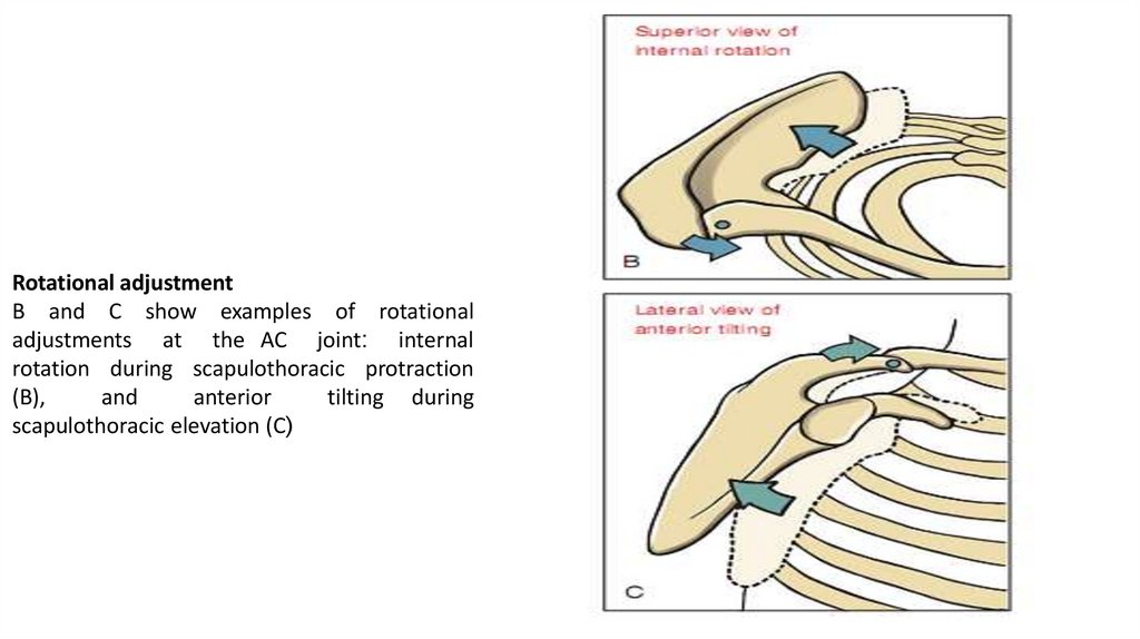
Rotational adjustmentB and C show examples of rotational
adjustments at the AC joint: internal
rotation during scapulothoracic protraction
(B),
and
anterior
tilting during
scapulothoracic elevation (C(
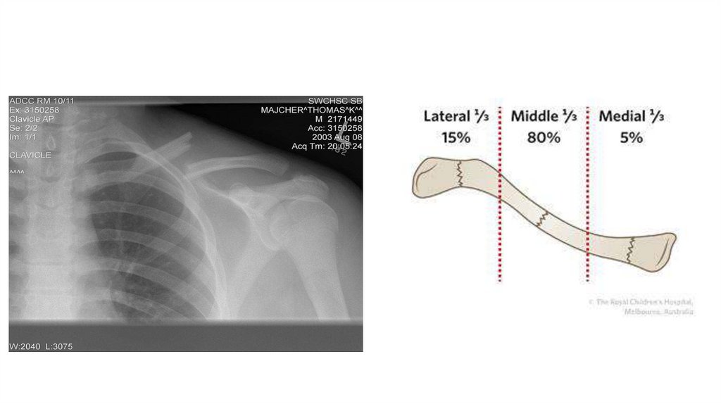
12. Clavicle Fractures
ClassificationThere is more than one accepted classification for clavicle fractures.
The following is Craig's classification:
1. Group I-fracture of the middle one third (most clavicular fractures
are group I fractures)
2. Group II-fracture of the lateral or distal one third
3. Group III-fracture of the medial one third
13.
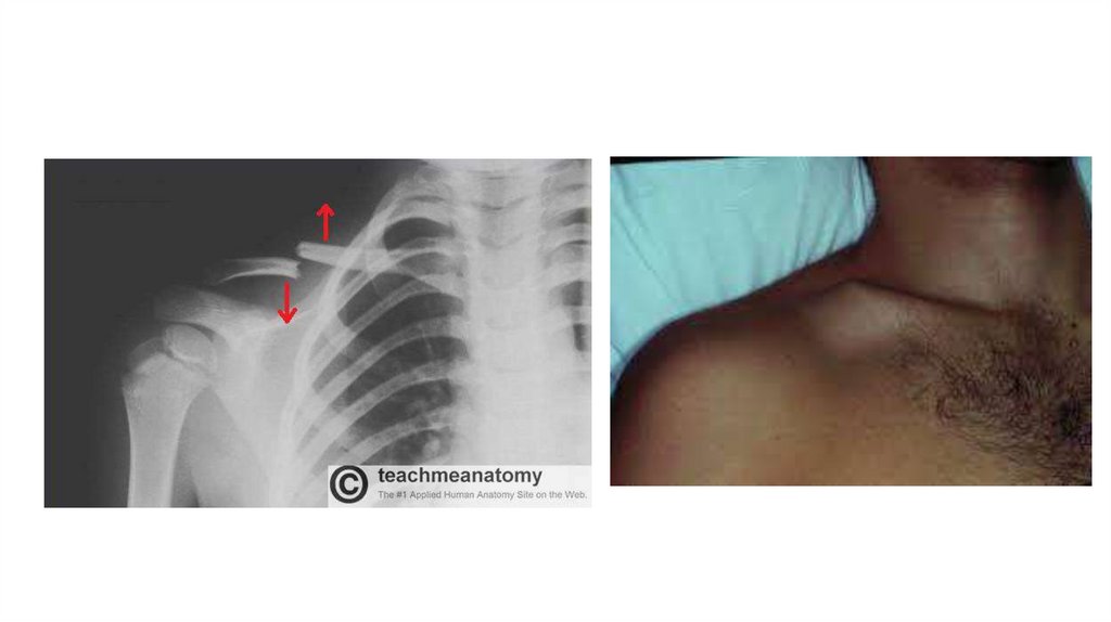
14. Mechanism of Injury
• Most clavicle fractures are caused by a fallor other direct trauma to the shoulder
• Falls on an outstretched hand (FOOSH),
although commonly cited, account for a
smaller percentage of clavicle fractures.
15.
16. Treatment Goals
1. Range of MotionRestore and improve the range of motion of the shoulder girdle.
2. Muscle Strength
Improve the strength of the following muscles:
Sternocleidomastoid (neck rotation)
Pectoralis major (arm adduction)
Deltoid (arm abduction)
17.
3. Functional GoalsImprove and restore the function of the shoulder for
activities of daily living and vocational and sports activities.
• Expected Time of Bone Healing 6 to 12 weeks.
• Expected Duration of Rehabilitation 10 to 12 weeks.
18. Treatment methods
Sling or Supportive Immobilization• This is the method of choice for most clavicle fractures. Many
comparisons of plain sling treatment to figure-of-eight bracing in
adults have shown no difference.
Open Reduction and Internal Fixation
• Open reduction and internal fixation is the method of choice for open
fractures
19. Arm sling
Figure-of-eight bracing20. TREATMENT
Treatment: Early to Immediate (Day of Injury to One Week)• BONE HEALING
• Stability at fracture site: None.
• Stage of bone healing: Inflammatory phase.
• X-ray: No callus.
21. Physical Examination
1. Check for capillary refill.2. Sensory evaluation.
3. The active and passive range of motion of the affected extremity,
including the elbow, wrist, and digits.
4. Evaluate any incision site for drainage, erythema.
5. Evaluate the patient's neurovascular status with a thorough brachial
plexus assessment.
6. Check the sling for proper fit and padding at the axillary area and
back of the neck
22. Prescription
• Precautions: Shoulder is held in adduction and internal rotation. Elbow ismaintained at 90 degrees of flexion.
• Range of Motion: No range of motion to the shoulder. Full, active range
of motion is encouraged to the wrist, hand, and digits.
• Muscle Strength: No strengthening exercises to the shoulder, Begin
isometric exercises to elbow arm and wrist 3 to 4 days after the fracture,
once the pain subsides.
23. Prescription
• Functional Activities:Personal hygiene The uninvolved extremity is used in self-care and
personal hygiene.
Bed mobility: The patient is instructed to roll over to the unaffected
side to come to a sitting position in bed.
• Weight bearing : None.
24. Treatment: 2 to 4 Weeks
• Stability at fracture site: None to minimal• Stage of bone healing: begin to reparative phase.
• X-ray: No to early callus. (Visible fracture line)
25. Prescription
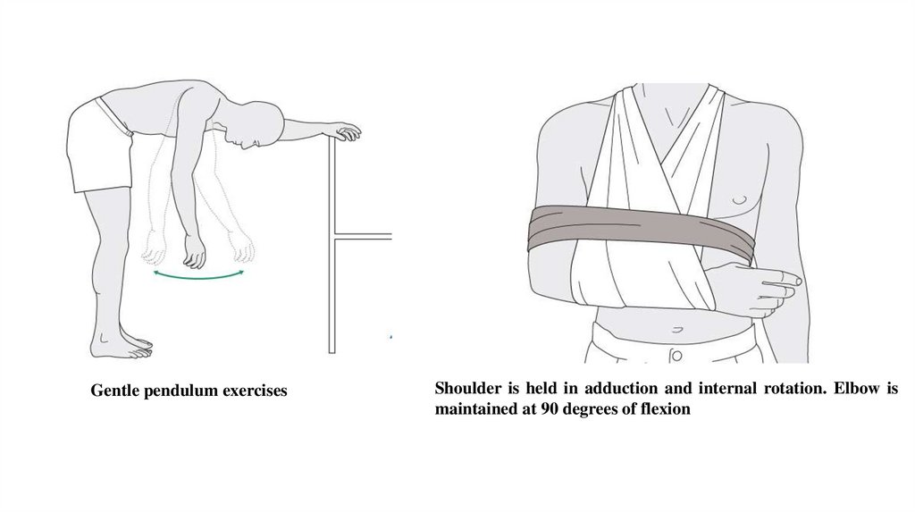
• Precautions: The same as previous weeks.• Range of Motion: Gentle pendulum exercises to the shoulder in the
sling as pain permits.
• Muscle Strength: No strengthening exercises to the shoulder. Start
gentle isometric exercises to the deltoid.
• Functional Activities: The same as previous weeks.
• Weight bearing: None.
26.
Gentle pendulum exercisesShoulder is held in adduction and internal rotation. Elbow is
maintained at 90 degrees of flexion
27. Treatment: 4 to 6 weeks
BONE HEALING• Stability at fracture site: with bridging callus the fracture is usually
stable.
• Stage of bone healing: Reparative phase.
• X-ray: Bridging callus is visible.
28. Prescription
• Precautions: At the end of 6 weeks, once there is good callusformation and the fracture site is stable, the sling or brace is removed
with limitation of abduction.
• Range of Motion: At the end of 6 weeks, gentle active assisted range
of motion to the shoulder is allowed. Abduction is limited to 80
degrees.
29. Prescription
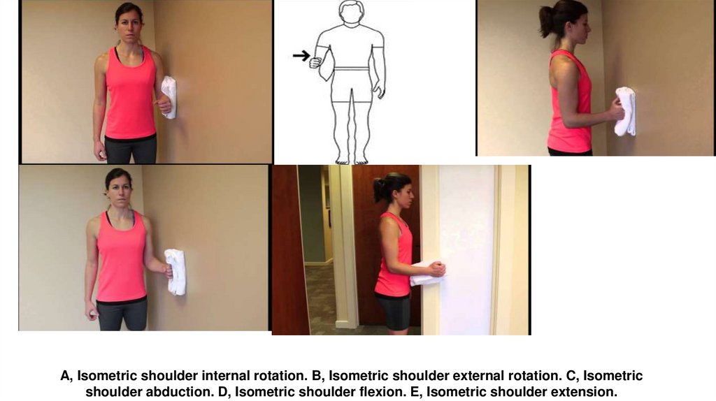
• Muscle Strength: Start isometric exercises to the rotator cuff anddeltoids.
• Functional Activities: The patient uses the affected extremity for some
self-care and personal hygiene.
• Weight bearing: None.
30.
A, Isometric shoulder internal rotation. B, Isometric shoulder external rotation. C, Isometricshoulder abduction. D, Isometric shoulder flexion. E, Isometric shoulder extension.
31. Treatment: 6 to 8 Weeks
BONE HEALING• Stability at fracture site: with bridging callus the fracture is usually
stable.
• Stage of bone healing: Reparative phase.
• X-ray: Bridging callus more apparent.
32.
Prescription• Precautions: Avoid contact sports.
• Range of Motion: active range of motion in all planes.
• Muscle Strength: Resistive exercises to the shoulder girdle muscles.
• Functional Activities: The patient uses the affected extremity for
personal hygiene, self-care, stabilization, and light activities.
• Weight bearing: Gradual weight bearing is allowed (when pushing off
from a chair or bed or using axillary crutches or a cane).
33. Treatment: 8 to 12 Weeks
BONE HEALING• Stability at fracture site: Stable.
• Stage of bone healing: Remodeling phase.
• X-ray: Bridging callus is very visible.
• N.B frequently, it takes years for the large callus to remodel and be less
visible cosmetically.
34.
Prescription• Precautions: Contact sports should be avoided for approximately 2
months.
• Range of Motion: Full ROM with abduction is encouraged.
• Muscle Strength: The resistance is gradually increased.
• Functional Activities: The involved extremity is used in self-care and
functional activities.
• Weight bearing: Full weight bearing.
35. Proximal Humeral Fractures
• DefinitionFractures of the proximal end of the humerus involve the humeral
head, anatomic neck, and surgical neck of the humerus.
Neer's classification system categorizes these fractures as one-, two-,
three-, or four-part fractures based on the displacement and
angulation of the parts, which are:
the head, shaft, greater tuberosity, and lesser tuberosity in the
proximal humerus.
36.
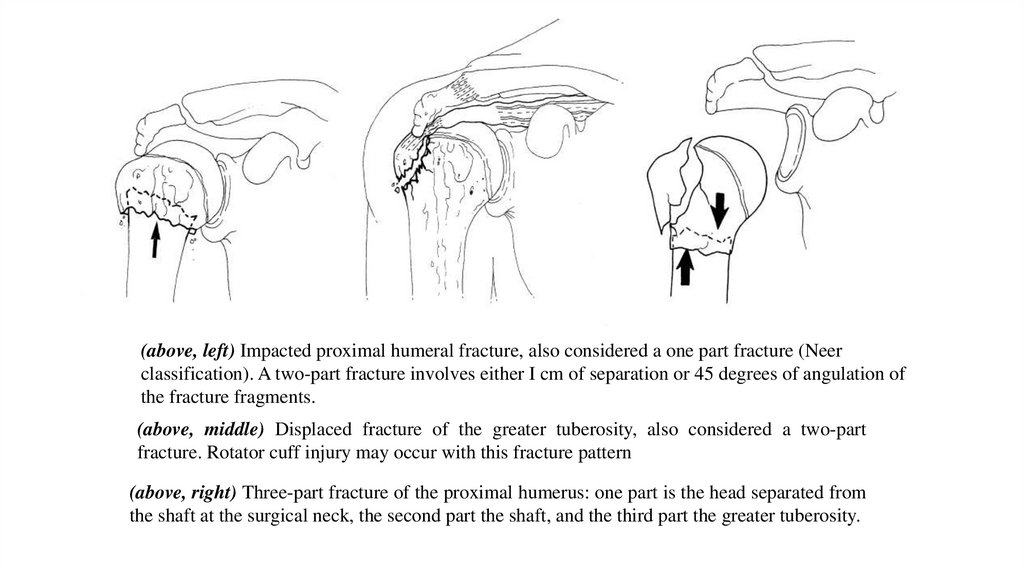
(above, left) Impacted proximal humeral fracture, also considered a one part fracture (Neerclassification). A two-part fracture involves either I cm of separation or 45 degrees of angulation of
the fracture fragments.
(above, middle) Displaced fracture of the greater tuberosity, also considered a two-part
fracture. Rotator cuff injury may occur with this fracture pattern
(above, right) Three-part fracture of the proximal humerus: one part is the head separated from
the shaft at the surgical neck, the second part the shaft, and the third part the greater tuberosity.
37.
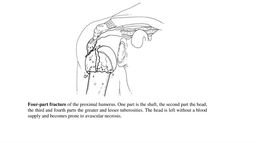
Four-part fracture of the proximal humerus. One part is the shaft, the second part the head,the third and fourth parts the greater and lesser tuberosities. The head is left without a blood
supply and becomes prone to avascular necrosis.
38. X-ray
39.
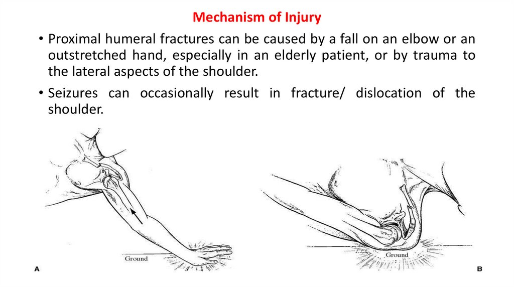
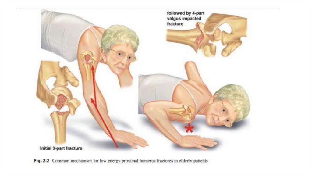
Mechanism of Injury• Proximal humeral fractures can be caused by a fall on an elbow or an
outstretched hand, especially in an elderly patient, or by trauma to
the lateral aspects of the shoulder.
• Seizures can occasionally result in fracture/ dislocation of the
shoulder.
40.
41. Treatment Goals
Rehabilitation Objectives1-Restore the full range of motion of the shoulder in all planes.
Frequently, there may be residual loss of range of motion secondary to
the fracture
2-Improve the strength of the shoulder muscles , especially of the
deltoid muscles.
3-Improve and restore the function of the shoulder for activities of
daily living and sports activities.
42. Treatment Methods
1. SlingIndications: undisplaced, impacted, or minimally displaced fractures usually
are immobilized for 2 to 3 weeks until the patient's pain subsides.
85 % of proximal humeral fractures are minimally displaced.
2. Open Reduction and Internal Fixation
Indications: two- and three-part fractures and those that may also require
repair of the rotator cuff
3. External Fixator
Indications: used for open and severely comminuted fractures.
43.
• Expected Time of Bone Healing6 to 8 weeks.
• Expected Duration of Rehabilitation
12 weeks to 1 year.
• Associated Injury
Rotator cuff Tears are associated with displacement of either tuberosity
and require repair .
Neurovascular Injuries
axillary nerve or posterior cord of the brachial plexus
44. TREATMENT
Treatment: Early (Day of Injury to One Week)BONE HEALING
Stability at fracture site: None.
Stage of bone healing: Inflammatory phase.
X-ray: No callus.
45.
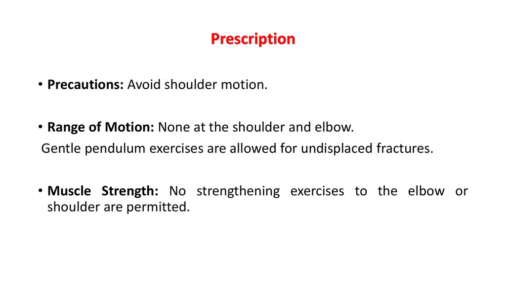
Prescription• Precautions: Avoid shoulder motion.
• Range of Motion: None at the shoulder and elbow.
Gentle pendulum exercises are allowed for undisplaced fractures.
• Muscle Strength: No strengthening exercises to the elbow or
shoulder are permitted.
46. Prescription
• Functional Activities: One-handed activities with the uninvolvedextremity. The patient needs assistance in dressing, grooming, and
preparing meals.
• Weight Bearing: None on affected extremity.
47. Treatment: 2 to 4 Weeks
BONE HEALINGStability at fracture site: None to minimal.
Stage of bone healing: Beginning of reparative phase.
X-ray: No callus; fracture line is still visible.
Prescription
Precautions: Avoid internal/external rotation of the shoulder because
they displace the fracture.
48.
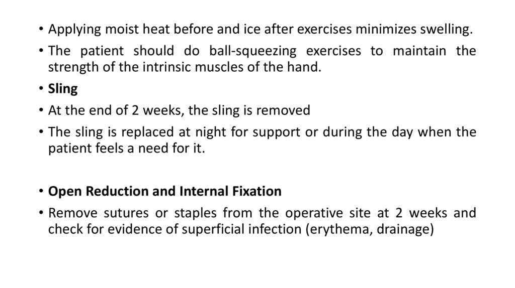
• Applying moist heat before and ice after exercises minimizes swelling.• The patient should do ball-squeezing exercises to maintain the
strength of the intrinsic muscles of the hand.
• Sling
• At the end of 2 weeks, the sling is removed
• The sling is replaced at night for support or during the day when the
patient feels a need for it.
• Open Reduction and Internal Fixation
• Remove sutures or staples from the operative site at 2 weeks and
check for evidence of superficial infection (erythema, drainage)
49.
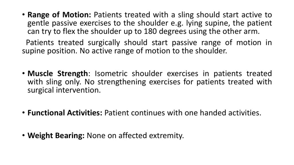
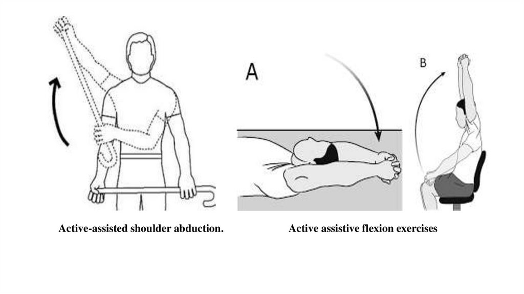
• Range of Motion: Patients treated with a sling should start active togentle passive exercises to the shoulder e.g. lying supine, the patient
can try to flex the shoulder up to 180 degrees using the other arm.
Patients treated surgically should start passive range of motion in
supine position. No active range of motion to the shoulder.
• Muscle Strength: Isometric shoulder exercises in patients treated
with sling only. No strengthening exercises for patients treated with
surgical intervention.
• Functional Activities: Patient continues with one handed activities.
• Weight Bearing: None on affected extremity.
50.
Active-assisted shoulder abduction.Active assistive flexion exercises
51. Treatment: 4 to 6 Weeks
BONE HEALINGStability at fracture site: With bridging callus, the fracture is usually
stable.
Stage of bone healing: reparative phase.
X-ray: Bridging callus is visible.
Prescription
Precautions: Do not apply force in attempting to regain the full range of
motion.
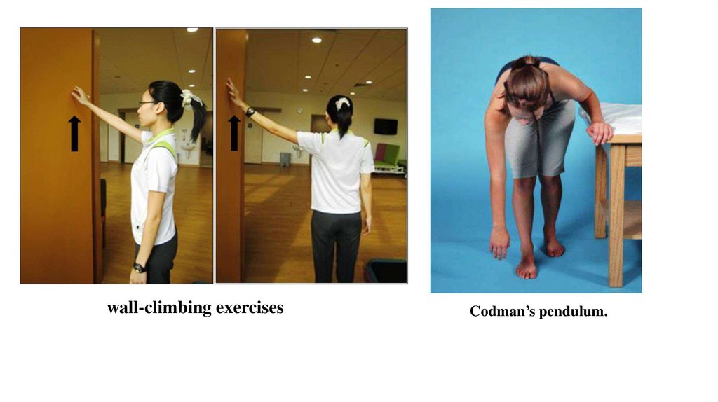
Range of Motion: Shoulder-limited range Flexion/abduction up to 100 to
110 degrees e.g. wall-climbing exercises (fingers against the wall and
reaching up)
52.
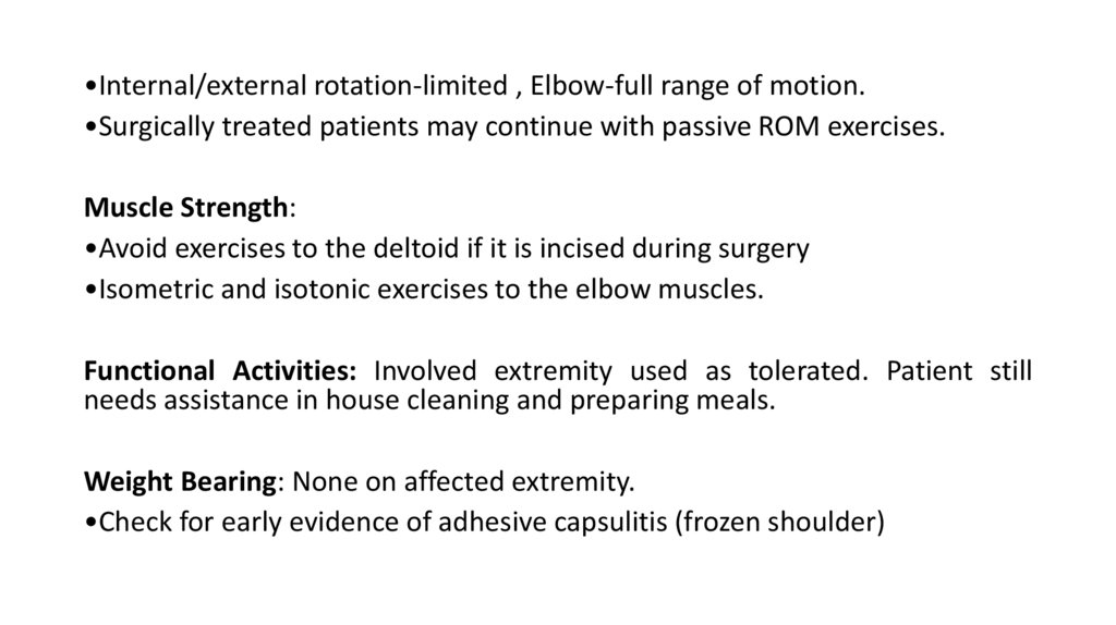
•Internal/external rotation-limited , Elbow-full range of motion.•Surgically treated patients may continue with passive ROM exercises.
Muscle Strength:
•Avoid exercises to the deltoid if it is incised during surgery
•Isometric and isotonic exercises to the elbow muscles.
Functional Activities: Involved extremity used as tolerated. Patient still
needs assistance in house cleaning and preparing meals.
Weight Bearing: None on affected extremity.
•Check for early evidence of adhesive capsulitis (frozen shoulder)
53.
wall-climbing exercisesCodman’s pendulum.
54. Treatment: 6 to 8 Weeks
BONE HEALINGStability at fracture site: With bridging callus, the fracture is usually
stable.
Stage of bone healing: reparative phase.
X-ray: Bridging callus is visible.
55.
PrescriptionPrecautions: Avoid forced range of motion.
Range of Motion: Active, active-assistive, and passive range of motion
to the shoulder and elbow in all planes, to tolerance.
Muscle Strength:
•Continue isometric exercises to the shoulder , and isotonic exercises
to the elbow.
•Start progressive resistive exercises for patients treated with a sling.
Functional Activities: The involved extremity is used for self-care and
feeding.
Weight Bearing: Weight bearing as tolerated
56.
Treatment: 8 to 12 WeeksBONE HEALING
Stability at fracture site: stable.
Stage of bone healing: Remodeling phase.
X-ray: Abundant callus; fracture line begins to disappear.
Prescription
Muscle Strength:
•Resistive exercises to the shoulder with gradual increases in weights.
Weight Bearing: full Weight bearing.
57.
References1. David J. Magee, Pathology and Intervention in Musculoskeletal
Rehabilitation , 2nd ed. 2016
2. Rehabilitation for the Postsurgical Orthopedic Patient, 3rd Edition.
2013
3. Treatment and Rehabilitation of Fractures. 2000
4. David J. Magee , Orthopedic Physical Assessments Atlas And Video:
Selected Special Testes and Movements , 5th ed. 2011
5. James Wyss, Therapeutic programs for musculoskeletal disorders
.2013