


 Медицина
Похожие презентации:


Acute myocardial infarction
1.
HISTORY41-year-old man.
CHIEF COMPLAINT: Chest pain of one hour duration.
PRESENT ILLNESS:
While playing racquetball, the patient developed
nausea and epigastric discomfort that increased over 30 minutes. His distress
became an intense substernal ache that radiated into the neck. Fire rescue
was called, and he was immediately transferred to a nearby emergency
department.
Question:
Based on the history, what is your diagnosis?
43-1
2.
Answer:Acute myocardial infarction is the most likely diagnosis.
Based on this history alone, electrocardiographic monitoring was initiated
immediately upon his admission to the emergency department. In addition,
pertinent laboratory studies were ordered stat, including an ECG, chest X ray
and blood for cardiac biomarkers.
Other causes of severe, prolonged chest pain that must be considered include
pericarditis, aortic dissection, pulmonary embolism, musculoskeletal diseases
and gastrointestinal lesions, e.g. esophagitis, cholecystitis and pancreatitis.
Proceed
43-2
3.
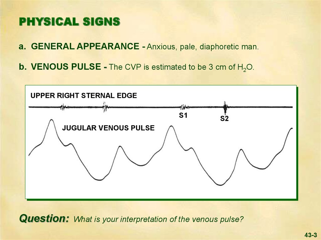
PHYSICAL SIGNSa. GENERAL APPEARANCE - Anxious, pale, diaphoretic man.
b. VENOUS PULSE - The CVP is estimated to be 3 cm of H2O.
UPPER RIGHT STERNAL EDGE
S1
S2
JUGULAR VENOUS PULSE
Question:
What is your interpretation of the venous pulse?
43-3
4.
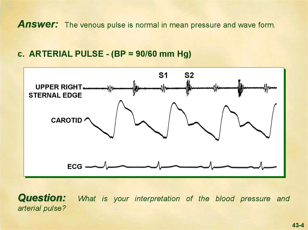
Answer:The venous pulse is normal in mean pressure and wave form.
c. ARTERIAL PULSE - (BP = 90/60 mm Hg)
S1
S2
UPPER RIGHT
STERNAL EDGE
CAROTID
ECG
Question:
What is your interpretation of the blood pressure and
arterial pulse?
43-4
5.
Answer:The blood pressure is mildly decreased, while the arterial pulse
contour is normal.
Parasympathetic overactivity may cause hypotension in this clinical setting, but
hypovolemia, arrhythmia, drug therapy and cardiac structural damage (e.g., left
ventricular dysfunction, right ventricular infarction, rupture of a papillary muscle,
septum or free wall) must also be considered.
Proceed
43-5
6.
d. PRECORDIAL MOVEMENTPHONO
UPPER RIGHT
STERNAL
EDGE
S1
S2
APEXCARDIOGRAM
Question:
How do you interpret the apical pulse?
43-6
7.
Answer:The apical impulse is normal
e. CARDIAC AUSCULTATION
UPPER LEFT STERNAL EDGE
.05 sec
INSPIRATION
S1
EXPIRATION
S1
S1
S1
S2
A2
P2
Question:
A2
S2
P2
How do you interpret the acoustic events at the upper left
sternal edge?
43-7
8.
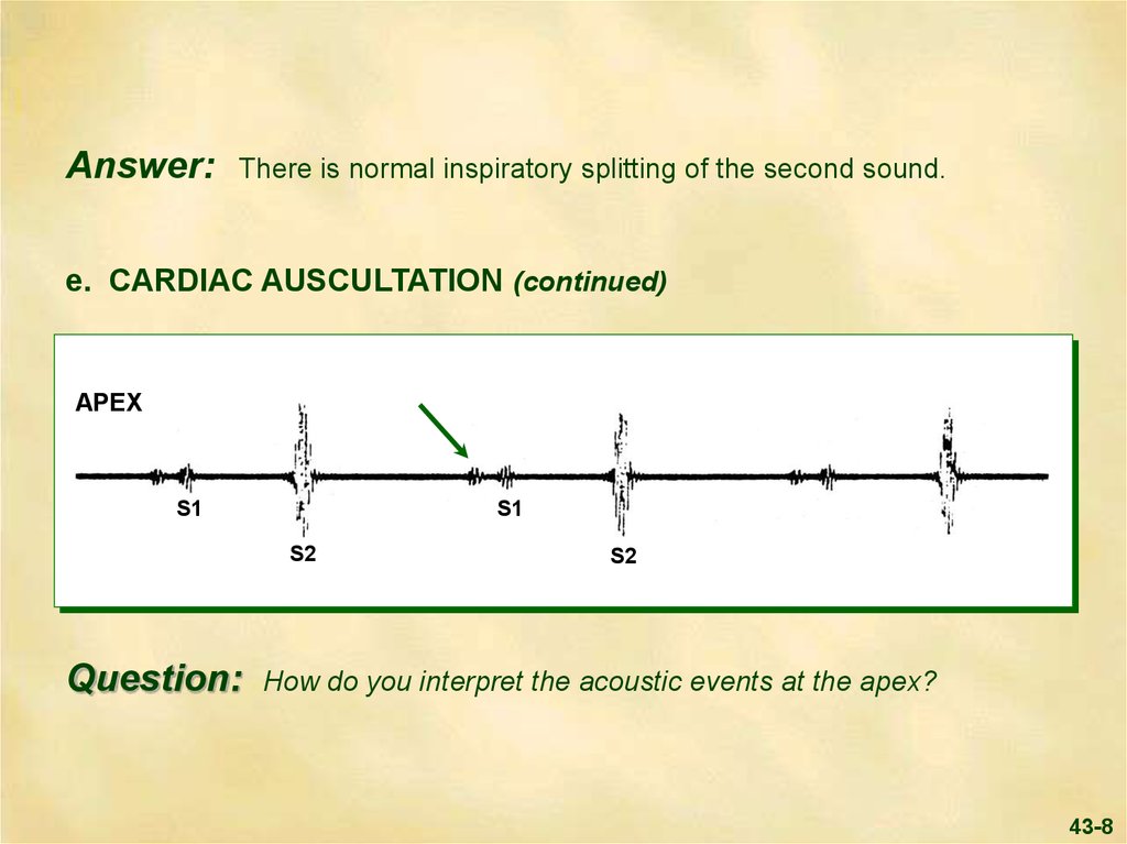
Answer:There is normal inspiratory splitting of the second sound.
e. CARDIAC AUSCULTATION (continued)
APEX
S1
S1
S2
Question:
S2
How do you interpret the acoustic events at the apex?
43-8
9.
Answer:The first heart sound at the apex is diminished in intensity. In this
clinical setting, factors such as reduced left ventricular contractility or early
closure of the mitral valve, as occurs with a prolonged PR interval, may
diminish the first sound.
The arrow points to the fourth heart sound heard at the apex. This reflects left
atrial contraction against a left ventricle with reduced compliance.
f. PULMONARY AUSCULTATION
Question:
How do you interpret the acoustic events in the pulmonary lung
fields?
Proceed
43-9
10.
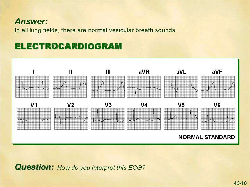
Answer:In all lung fields, there are normal vesicular breath sounds.
ELECTROCARDIOGRAM
I
II
III
aVR
aVL
aVF
V1
V2
V3
V4
V5
V6
NORMAL STANDARD
Question:
How do you interpret this ECG?
43-10
11.
Answer:The ECG shows marked ST segment elevation in the inferior
leads with lesser elevation in the lateral leads. There is ST segment
depression in leads I, aVL and V1-V3. This ECG is diagnostic of acute
inferolateral injury that almost always evolves to infarction.
This ECG also suggests that a significant amount of myocardium is in jeopardy,
as patients with anterior ST segment depression often have a larger infarction
than those with inferior ST segment elevation alone.
Note that the PR interval is prolonged, suggesting possible AV node ischemia
as is commonly seen in inferior wall infarction due to right coronary
artery obstruction.
Proceed
43-11
12.
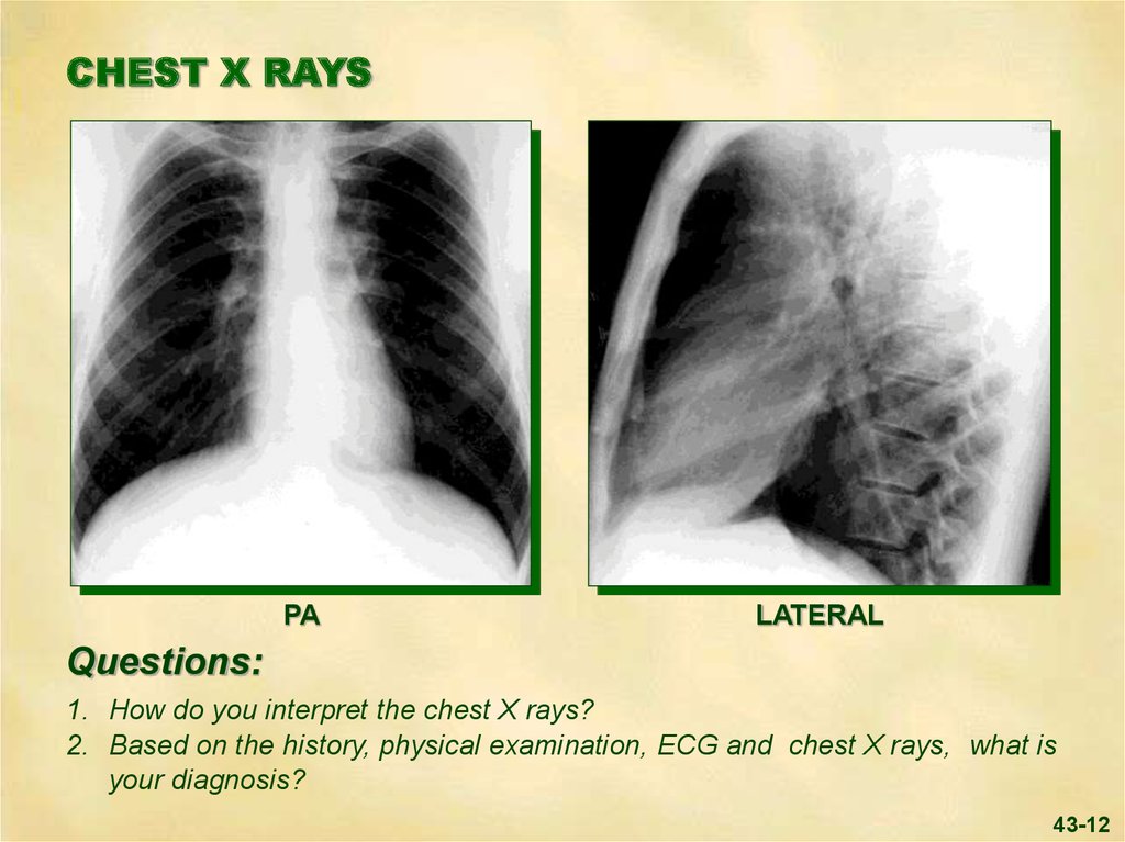
CHEST X RAYSPA
LATERAL
Questions:
1. How do you interpret the chest X rays?
2. Based on the history, physical examination, ECG and chest X rays, what is
your diagnosis?
43-12
13.
Answers:1. The chest X rays are normal.
2. Based on the history, physical examination, ECG and X rays, the diagnosis
is an evolving acute inferolateral wall myocardial infarction.
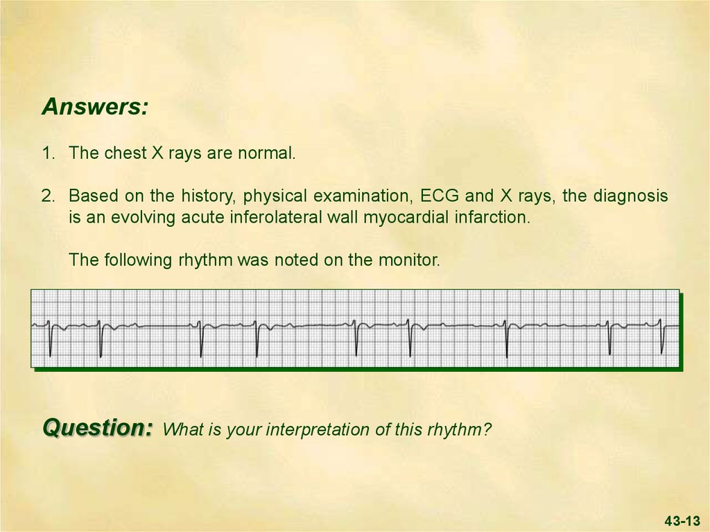
The following rhythm was noted on the monitor.
Question:
What is your interpretation of this rhythm?
43-13
14.
Answer:The rhythm strip shows sinus rhythm with Mobitz Type I second
degree A-V block ( Wenckebach) as evidenced by progressive lengthening of
the PR interval followed by a non-conducted sinus P wave.
A-V block is more common in inferior than in anterior wall infarction. The
spectrum from a prolonged PR interval, to second degree A-V block, to
complete A-V block may be seen. The conduction defect producing heart block
in patients with inferior infarction is usually located in the area of the A-V node,
rather than in the bundle of His or the bundle branches. The high incidence of
heart block in this setting is due to the fact that in 90% of patients the right
coronary artery supplies the A-V junction as well as the inferior wall. This type
of heart block is usually transient.
Question:
What is your plan of therapy for this patient?
43-14
15.
Answer:Thrombosis plays an important role in ST-elevation myocardial
infarction. Timely reperfusion of the occluded coronary artery can reduce
infarct size and decrease mortality. Reperfusion can be accomplished by
thrombolytic agents, percutaneous coronary intervention and coronary artery
bypass graft surgery.
Unless the patient is allergic to aspirin, it should be given as soon as possible.
If thrombolytics are used, the combination of an anti-platelet, anti-thrombin
(heparin), and fibrinolytic agent is necessary.
Nitroglycerin increases myocardial oxygen supply, especially when collaterals
are present, or if spasm is a component of coronary occlusion. Efforts should
also be made to decrease myocardial oxygen demand by use of a beta-blocker.
Proceed
43-15
16.
Answer(continued): If full catheterization facilities are available, urgent
study and percutaneous intervention is most often the treatment of choice.
Thrombolytic agents may also be an effective treatment if administered early in
the course of an acute myocardial infarction. Contraindications to such therapy
include any event or condition that predisposes to serious bleeding.
Because the catheterization lab was not immediate available, our patient was
treated within two hours of the onset of his symptoms with aspirin, a
thrombolytic agent and heparin. Beta-blockers were withheld because of his
slow heart rate.
Proceed
43-16
17.
LABORATORYMyocardial biomarkers ordered on admission confirmed the diagnosis of
infarction. Necrosis of myocardial tissue results in the release of intracellular
biomarkers into the blood. In this case, their transient rise was typical.
Creatine kinase isoenzyme (CK-MB) elevation begins approximately 4 hours
after symptoms of infarction, and in the absence of early coronary reperfusion
peaks at about 24 hours. Troponins I and T rise slightly later and remain
elevated longer.
Depending upon the thrombolytic agent used, appropriate clotting studies
should be carried out.
This patient’s routine blood work and clotting parameters were normal.
Proceed
43-17
18.
The patient was placed at bed rest in the CCU.Ninety minutes following thrombolytic therapy, the patient showed clinical
evidence that was consistent with reperfusion: his chest pain resolved, his
blood pressure rose to 120/80 mm Hg. An ECG taken at this time follows.
Proceed
43-18
19.
ELECTROCARDIOGRAMI
II
III
aVR
aVL
aVF
V1
V2
V3
V4
V5
V6
NORMAL STANDARD
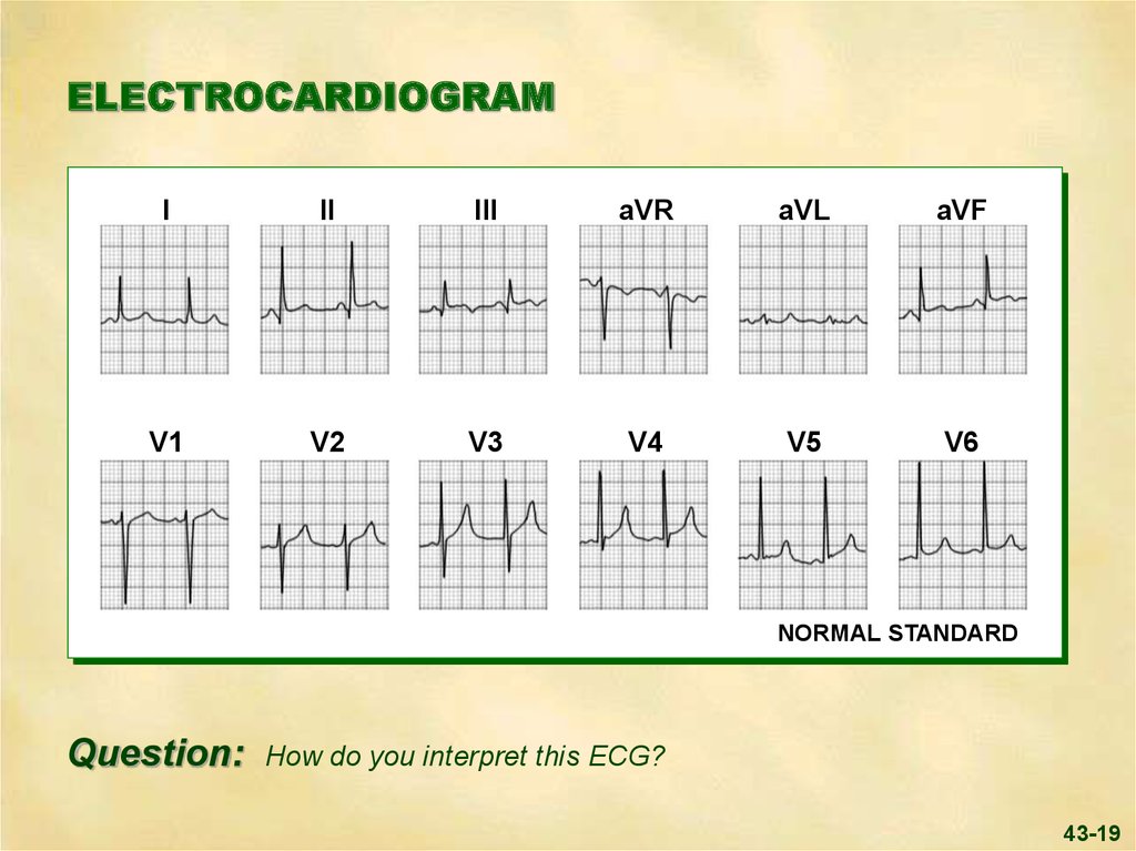
Question:
How do you interpret this ECG?
43-19
20.
Answer:Typical evolutionary changes of an acute inferior wall myocardial
infarction are present: Q waves and symmetrically inverted T waves are
seen in leads II, III and aVF, and the ST segments have returned to
the baseline. Reperfusion has accelerated these ECG changes in the
inferior wall and has resulted in the resolution of the other ST-T abnormalities
seen on the initial ECG. Note that his first degree heart block has resolved,
and the rhythm is sinus.
Proceed
43-20
21.
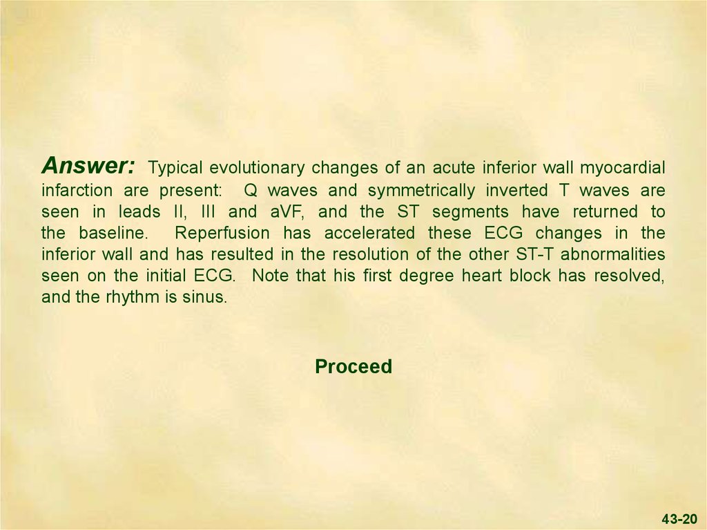
The patient remained clinically stable, although 30 minutes later the followingrhythm was seen on his monitor:
Question:
How would you interpret and treat this arrhythmia?
43-21
22.
Answer:There is a run of non-sustained ventricular tachycardia, i.e., three
or more ventricular beats in a row at a rate of over 100 but less than 30 beats.
Such “reperfusion ventricular arrhythmias” are frequently seen in this setting
and do not require therapy.
Proceed
43-22
23.
Two hours later the patient became cool and clammy, and his blood pressuredropped to 85/50 mm Hg. Because of its hypotensive effect, the nitroglycerin
infusion was discontinued.
The patient’s blood pressure rose to 105/70 mm Hg, but he was still clammy. A
bolus of IV saline was then given, as patients with acute myocardial infarction
may become hypovolemic, in part due to shifts in intravascular volume related
to catecholamine effect. The patient’s blood pressure then rose to
120/80 mm Hg, and he appeared alert and comfortable.
Over the next several days, the patient remained asymptomatic and stable and
his activity level gradually ambulated.
Proceed
43-23
24.
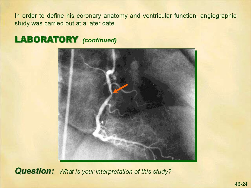
In order to define his coronary anatomy and ventricular function, angiographicstudy was carried out at a later date.
LABORATORY
Question:
(continued)
What is your interpretation of this study?
43-24
25.
Answer:The right coronary angiogram shows an isolated non-critical
stenosis (arrow) in the proximal right coronary artery. Additional views showed
an ulcerated plaque in this area, the probable site of a thrombus that was
present prior to thrombolytic therapy. The global ejection fraction was 55%
with mild inferior wall hypokinesis. This non-critical degree of obstruction
and the well preserved ejection fraction support the success of early
thrombolytic therapy in this case.
A Thallium stress test was carried out and demonstrated no significant
ischemia.
He was prescribed optimal therapy for secondary prevention that included
recommended life-style changes, aspirin, beta-blockers, ACE-inhibitors and
statins.
Proceed For Summary
43-25
26.
SUMMARYCoronary artery lesions range from the stable atheroma to complex lesions with
thrombotic occlusion. The primary event in most acute infarctions is ulceration
and/or rupture of an atherosclerotic plaque that becomes a nidus for platelet
aggregation and the development of a thrombus. The resulting abrupt
decrease in blood supply leads to cardiac tissue ischemia and necrosis.
There is greater myocardial salvage when efforts are directed towards the
prompt reopening of the occluded coronary artery. In order to salvage
jeopardized myocardium, therapy must be initiated before necrosis is complete.
The goal of early therapy is to decrease the size of the infarction and prevent
its complications. There is a significant difference in the ejection fraction and
overall mortality of patients in whom reperfusion was successful.
Proceed
43-26
27.
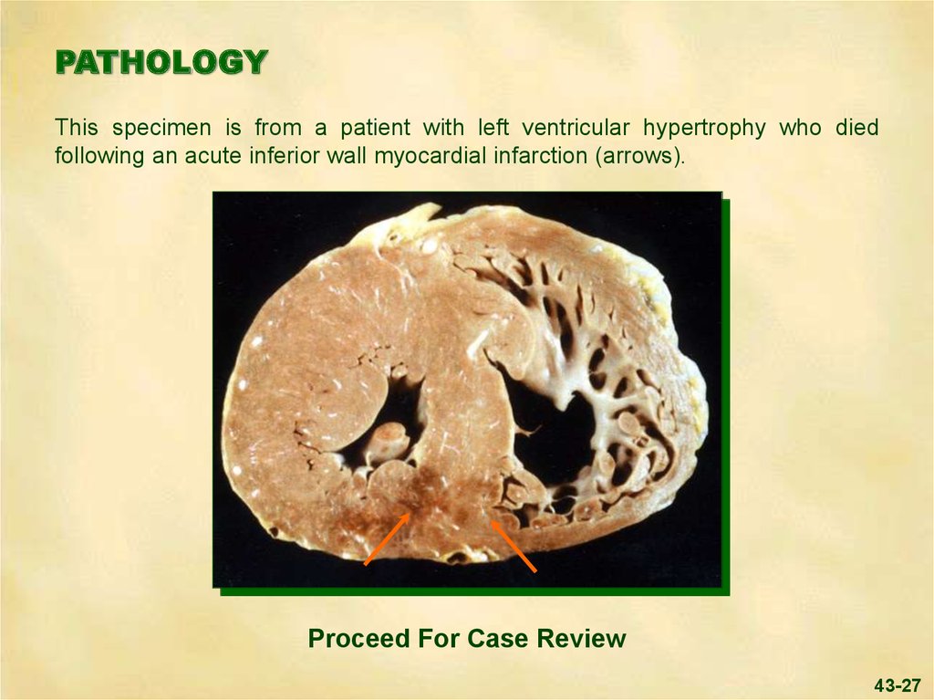
PATHOLOGYThis specimen is from a patient with left ventricular hypertrophy who died
following an acute inferior wall myocardial infarction (arrows).
Proceed For Case Review
43-27
28.
To Review This Case ofInferior Wall Myocardial Infarction:
The HISTORY is typical of an acute infarction, with the patient’s nausea
and epigastric discomfort suggesting the location may be inferior.
PHYSICAL SIGNS:
a. The GENERAL APPEARANCE reveals an anxious, pale,
man.
diaphoretic
b. The JUGULAR VENOUS PULSE is normal in mean pressure and
wave form.
c. The CAROTID ARTERIAL pulse contour is normal, but the blood
pressure is slightly decreased.
d. PRECORDIAL MOVEMENT reveals a normal early systolic impulse.
Proceed
43-28
29.
e. CARDIAC AUSCULTATION reveals an apical fourth heart sound,reflecting reduced left ventricular compliance. The intensity of S1 was
reduced, due to a prolonged PR interval.
f. PULMONARY AUSCULTATION reveals normal vesicular breath sounds
in all lung fields.
The CHEST
X RAYS are normal.
The ELECTROCARDIOGRAM shows first degree heart block and
ST elevation, typical of an inferolateral infarction.
Proceed
43-29
30.
LABORATORY STUDIES include cardiac biomarkers that confirmthe diagnosis of an evolving myocardial infarction. A rhythm strip revealed
transient Type I second degree A-V block.
Following thrombolysis,
non-sustained ventricular tachycardia occurred. Catheterization with
angiography revealed an isolated 50% right coronary obstruction, mild inferior
wall hypokinesis and a normal ejection fraction. An exercise Thallium study
was normal.
TREATMENT
with a thrombolytic agent resulted in reperfusion. His
second degree A-V block and ventricular tachycardia did not require treatment.
His hypotension responded well to discontinuation of his NTG and the
administration of fluids. The remainder of his hospital course was uneventful.
A secondary prevention program was instituted during his hospitalization.
43-30