


 Медицина
Похожие презентации:


Pneumonia
1.
Pneumonia2.
Definitions
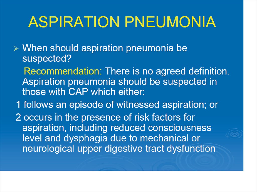
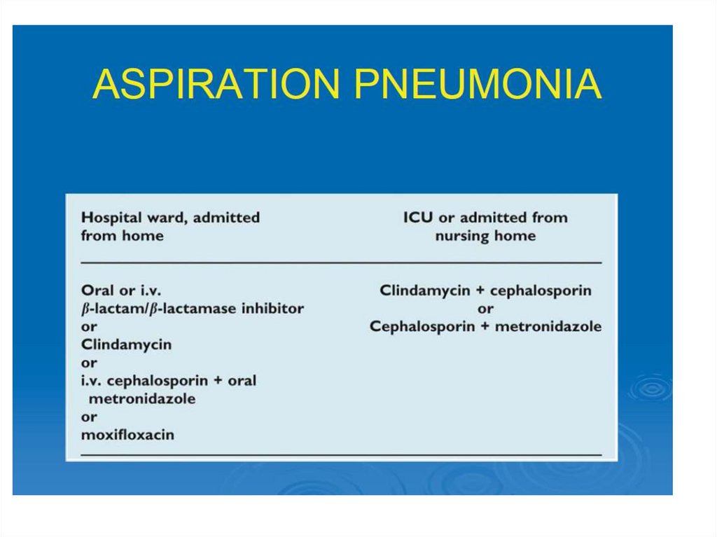
Ethiology (general), risk factors
Diagnosis criteria and evaluation
Peculiarities of the disease in different
causative agents
• Treatment
3.
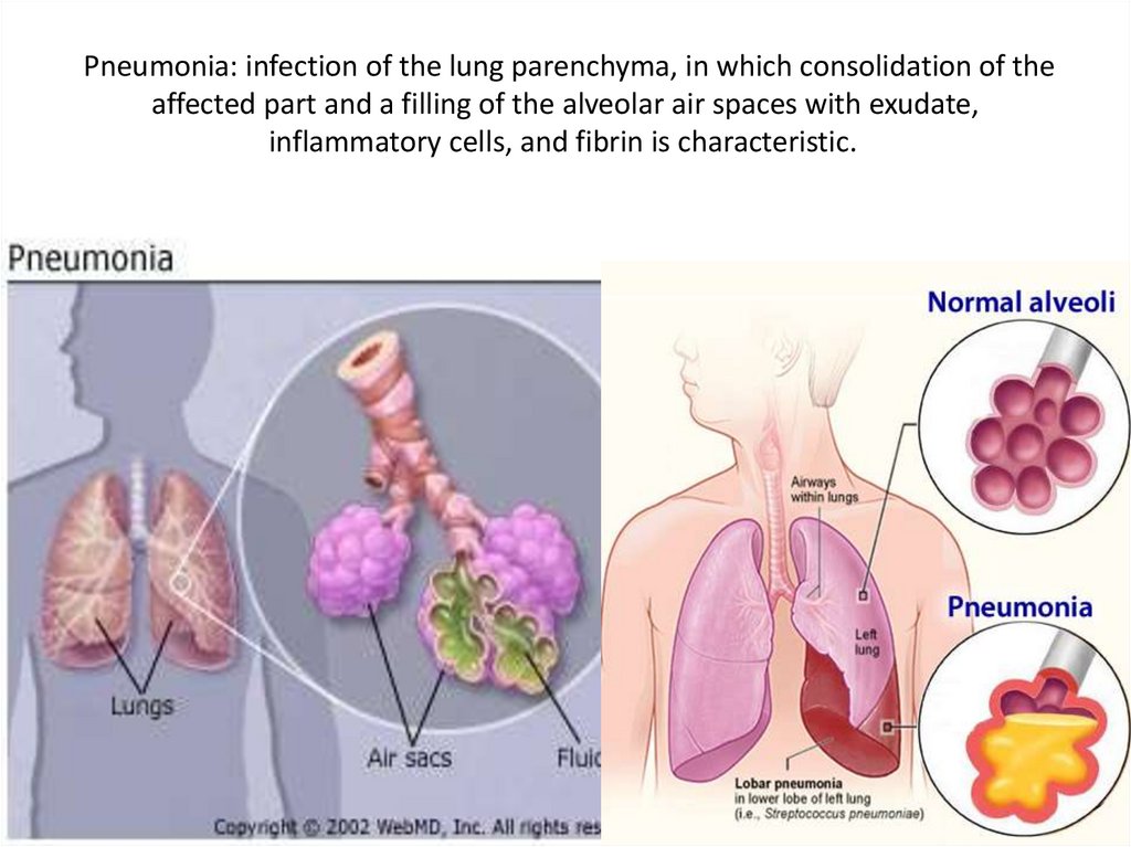
Pneumonia: infection of the lung parenchyma, in which consolidation of theaffected part and a filling of the alveolar air spaces with exudate,
inflammatory cells, and fibrin is characteristic.
4.
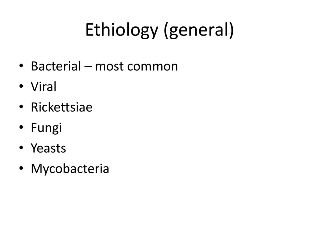
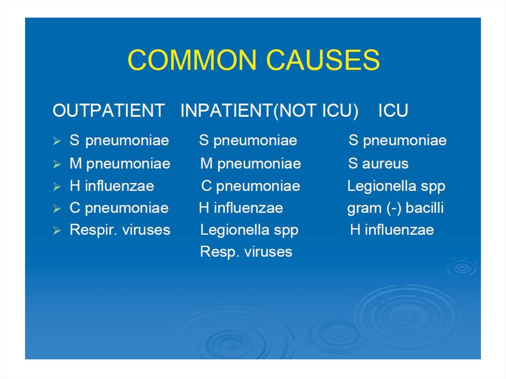
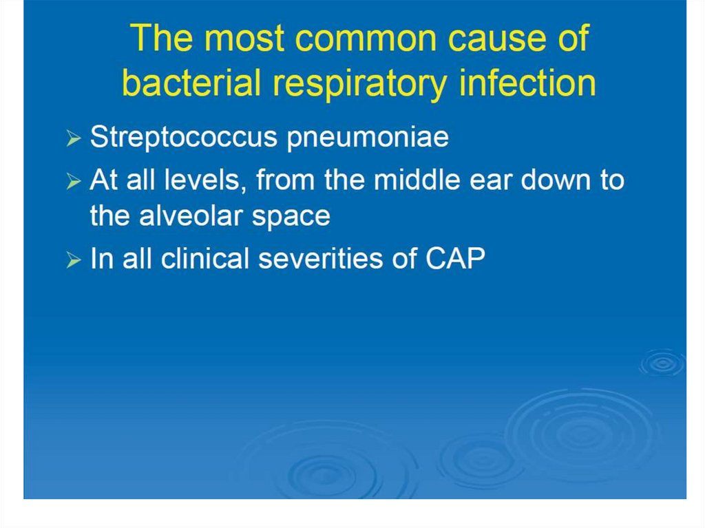
Ethiology (general)Bacterial – most common
Viral
Rickettsiae
Fungi
Yeasts
Mycobacteria
5.
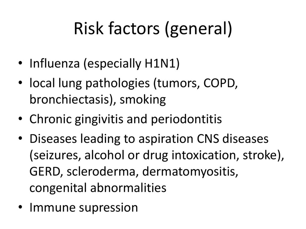
Risk factors (general)• Influenza (especially H1N1)
• local lung pathologies (tumors, COPD,
bronchiectasis), smoking
• Chronic gingivitis and periodontitis
• Diseases leading to aspiration CNS diseases
(seizures, alcohol or drug intoxication, stroke),
GERD, scleroderma, dermatomyositis,
congenital abnormalities
• Immune supression
6.
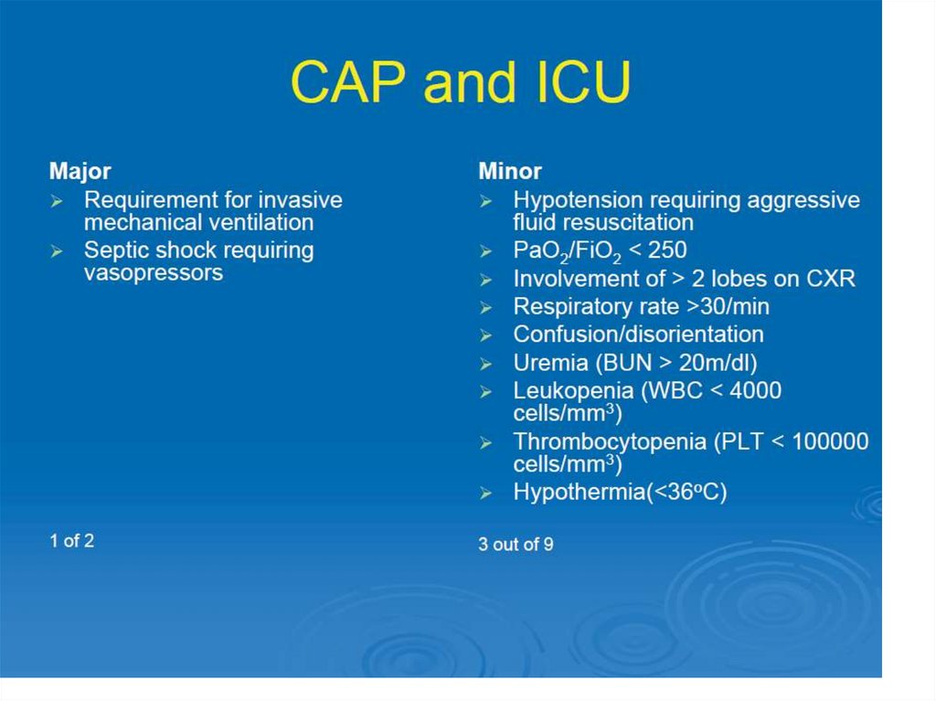
7.
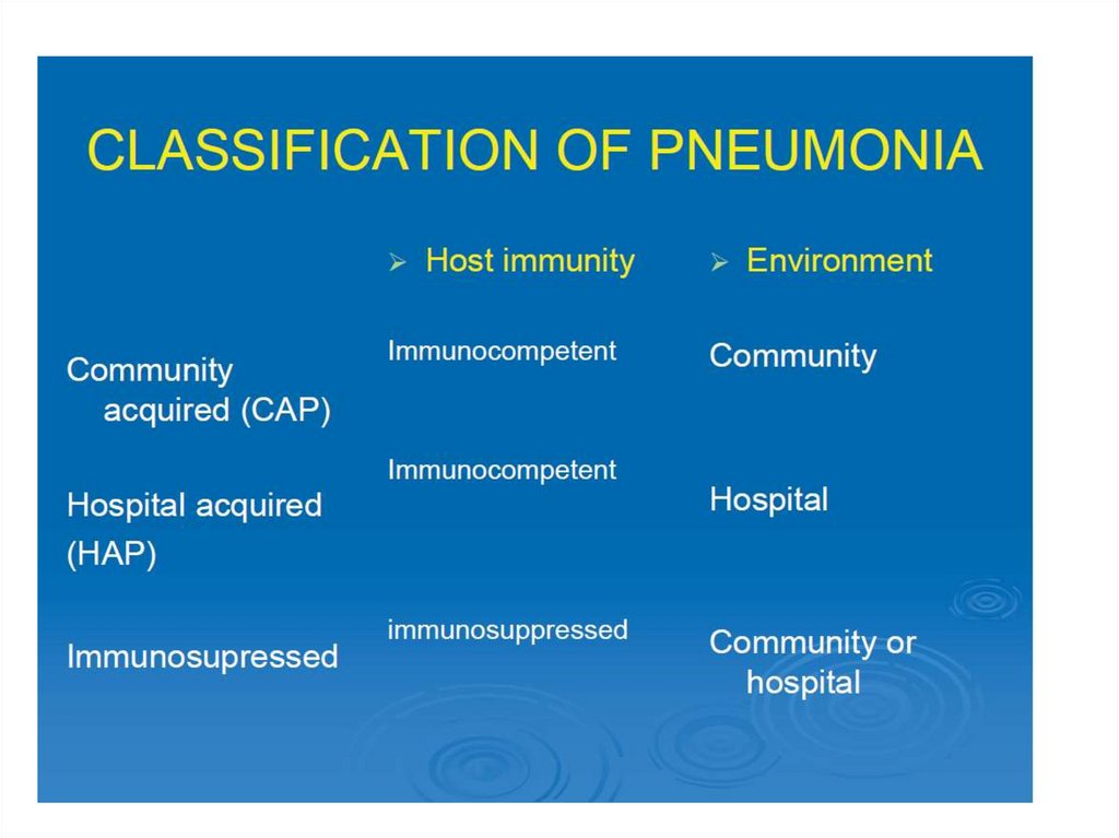
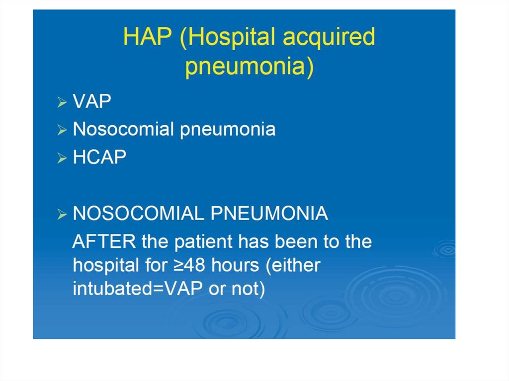
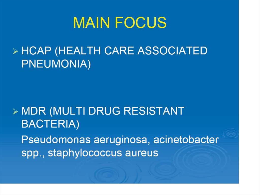
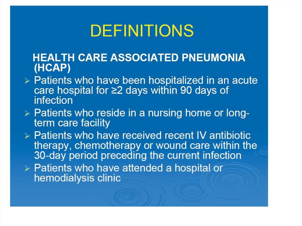
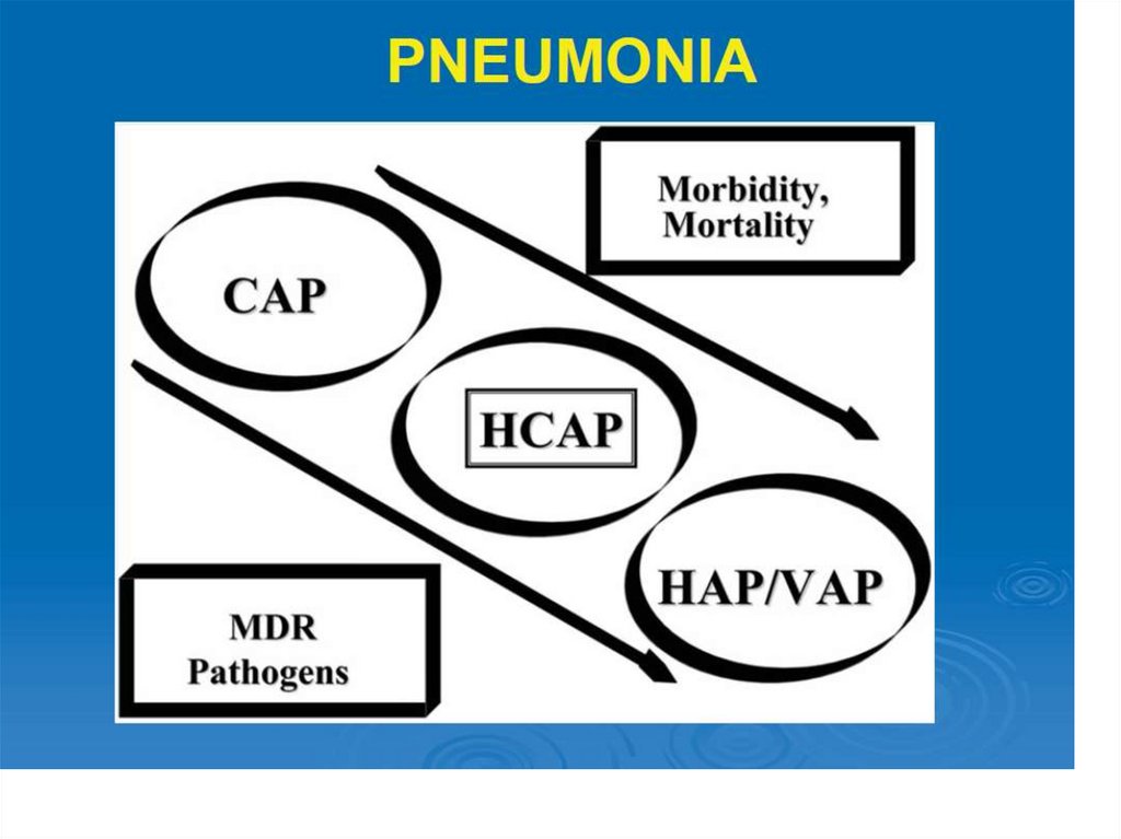
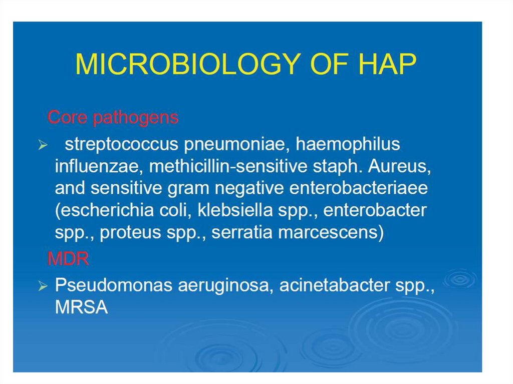
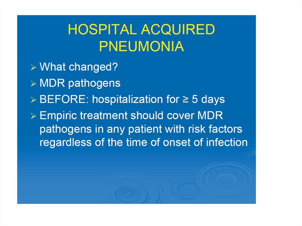
• CAP: Pneumonia not acquired in a hospital ora long-term care facility
• Hospital acquired pneumonia (with/without
multiple drug resistance risk factors):
- Healthcare associated pneumonia: other
healthcare facilities such as nursing homes,
dialysis centers, and outpatient clinics
- Hospital acquired pneumonia
- Ventilator associated pneumonia
8.
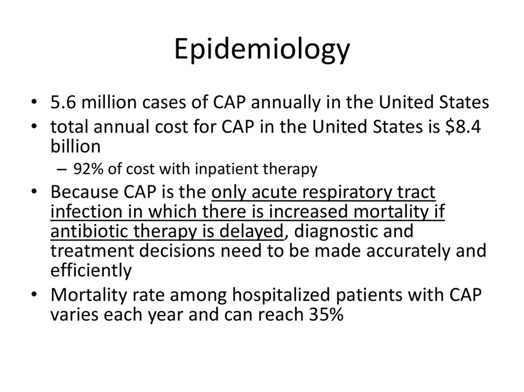
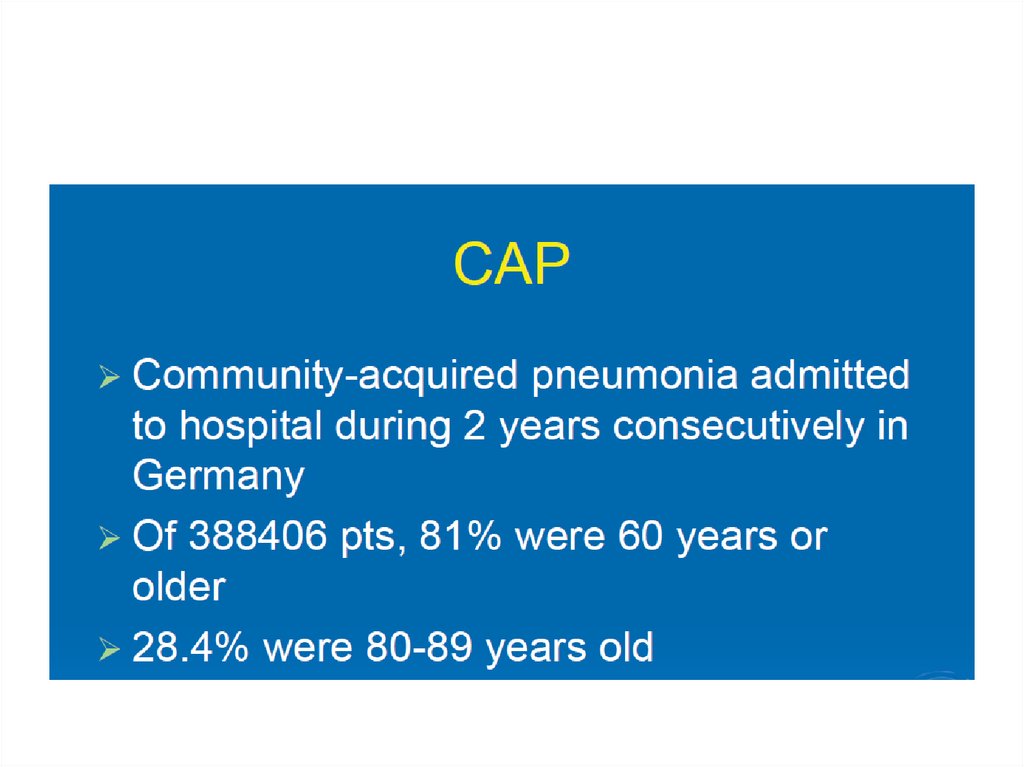
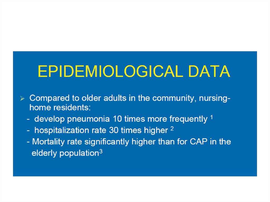
Epidemiology• 5.6 million cases of CAP annually in the United States
• total annual cost for CAP in the United States is $8.4
billion
– 92% of cost with inpatient therapy
• Because CAP is the only acute respiratory tract
infection in which there is increased mortality if
antibiotic therapy is delayed, diagnostic and
treatment decisions need to be made accurately and
efficiently
• Mortality rate among hospitalized patients with CAP
varies each year and can reach 35%
9.
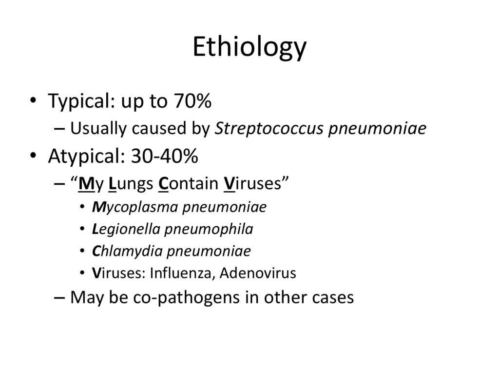
Ethiology• Typical: up to 70%
– Usually caused by Streptococcus pneumoniae
• Atypical: 30-40%
– “My Lungs Contain Viruses”
Mycoplasma pneumoniae
Legionella pneumophila
Chlamydia pneumoniae
Viruses: Influenza, Adenovirus
– May be co-pathogens in other cases
10.
11.
12.
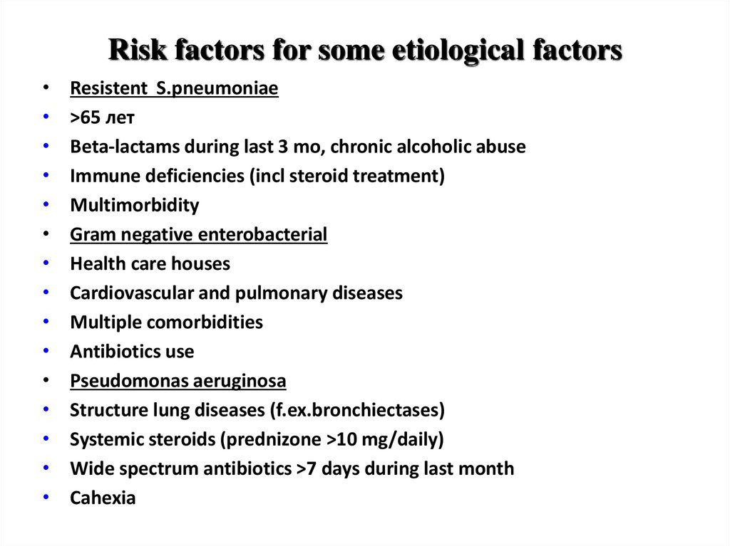
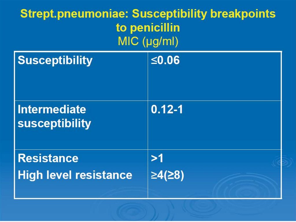
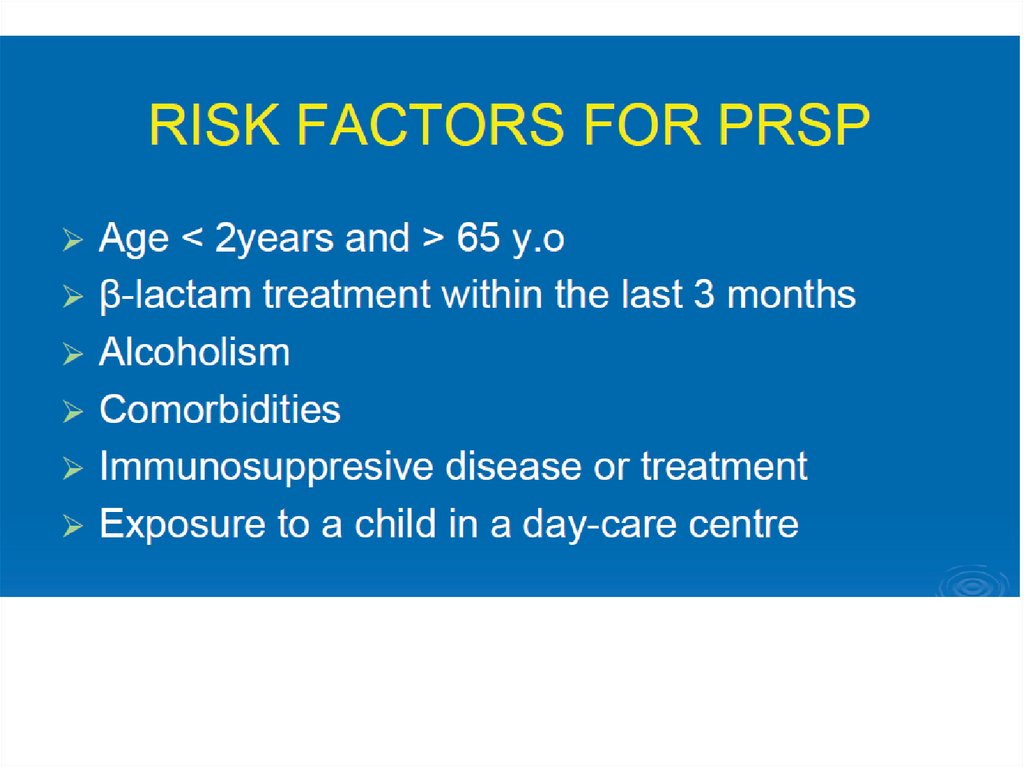
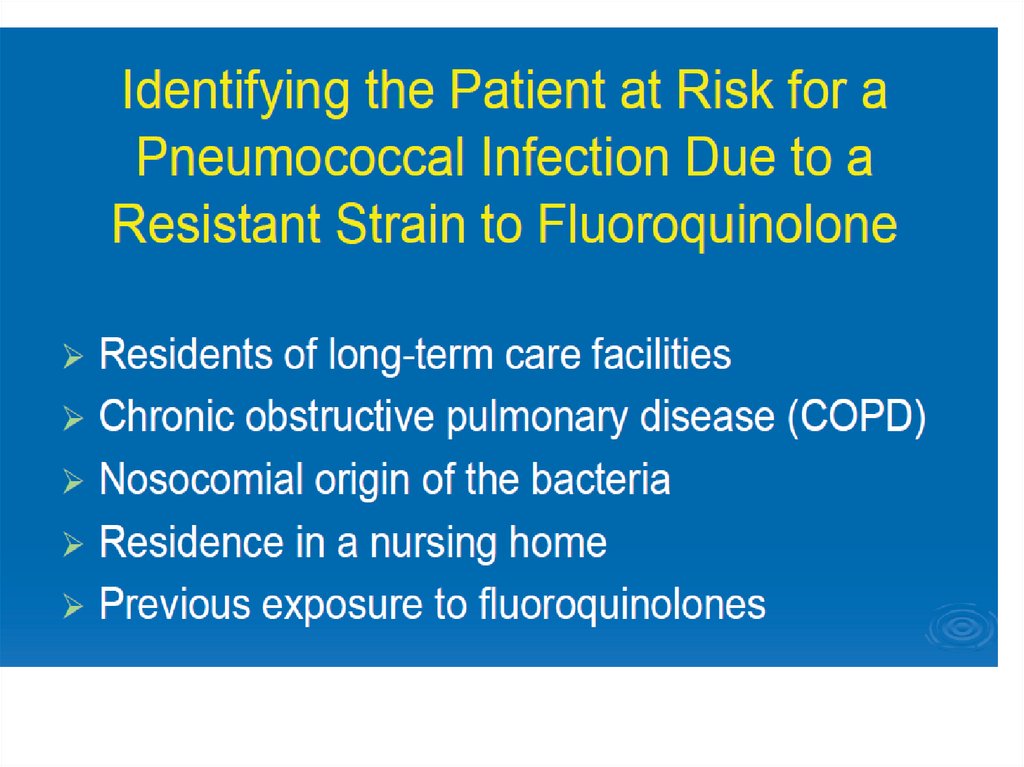
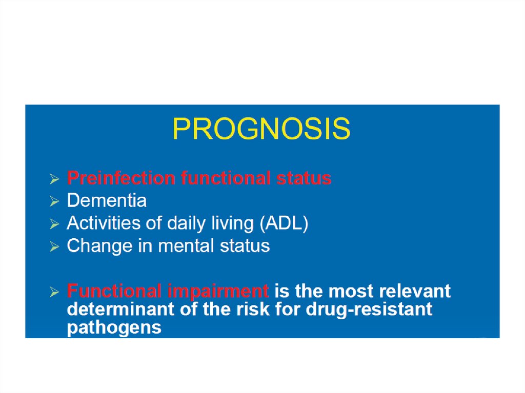
Risk factors for some etiological factorsResistent S.pneumoniae
>65 лет
Beta-lactams during last 3 mo, chronic alcoholic abuse
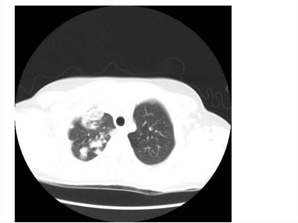
Immune deficiencies (incl steroid treatment)
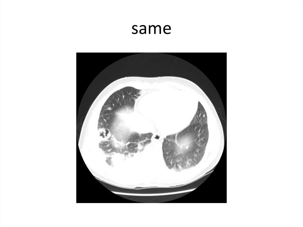
Multimorbidity
Gram negative enterobacterial
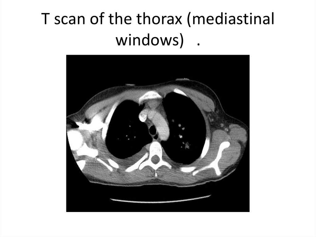
Health care houses
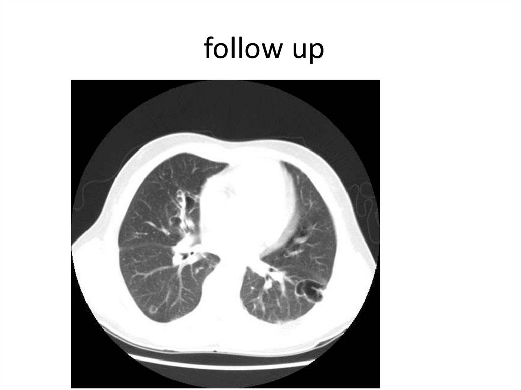
Cardiovascular and pulmonary diseases
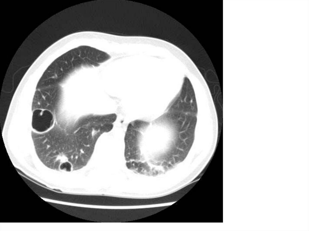
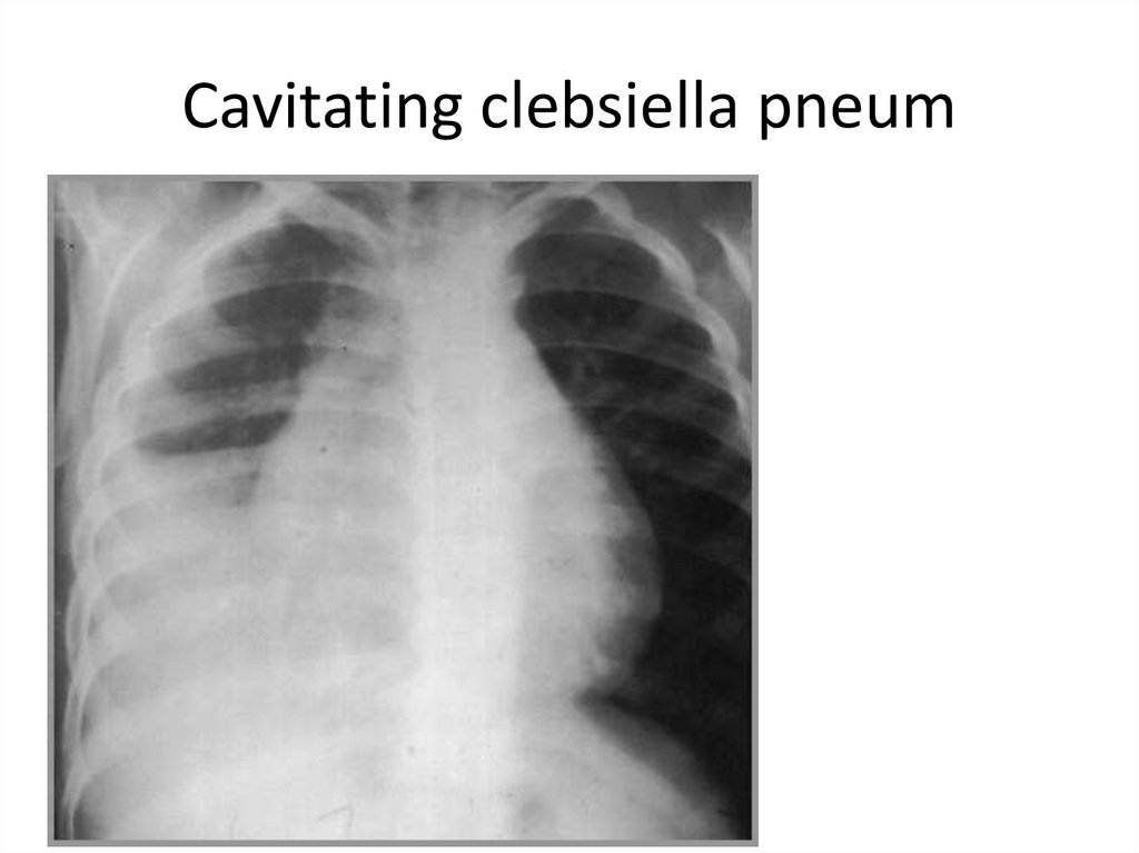
Multiple comorbidities
Antibiotics use
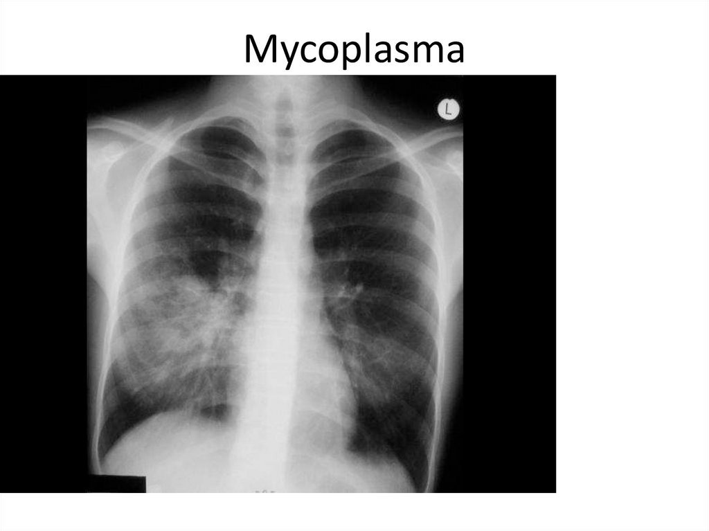
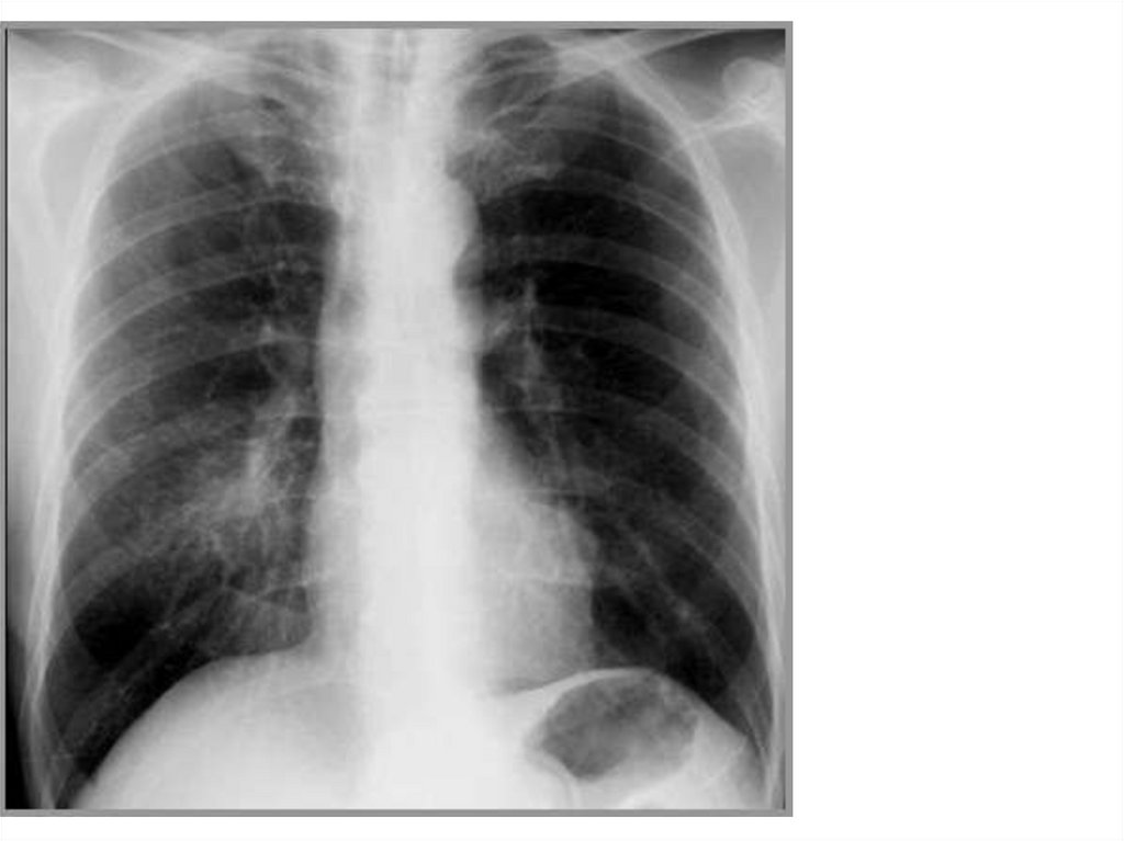
Pseudomonas aeruginosa
Structure lung diseases (f.ex.bronchiectases)
Systemic steroids (prednizone >10 mg/daily)
Wide spectrum antibiotics >7 days during last month
Cahexia
13.
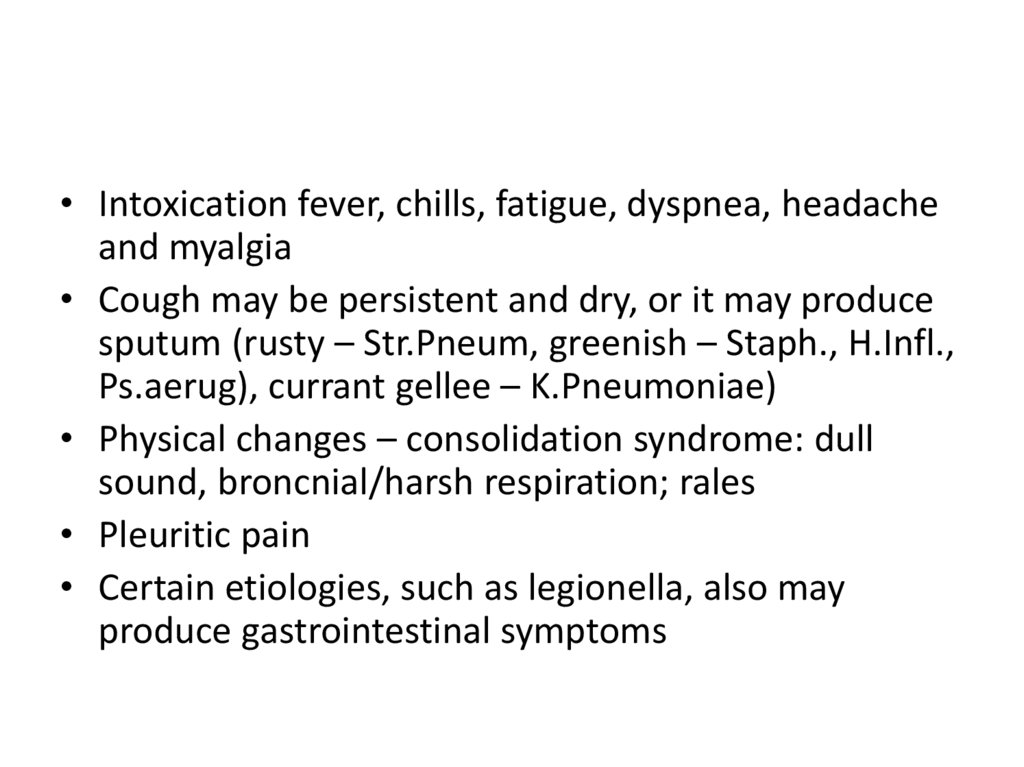
• Intoxication fever, chills, fatigue, dyspnea, headacheand myalgia
• Cough may be persistent and dry, or it may produce
sputum (rusty – Str.Pneum, greenish – Staph., H.Infl.,
Ps.aerug), currant gellee – K.Pneumoniae)
• Physical changes – consolidation syndrome: dull
sound, broncnial/harsh respiration; rales
• Pleuritic pain
• Certain etiologies, such as legionella, also may
produce gastrointestinal symptoms
14.
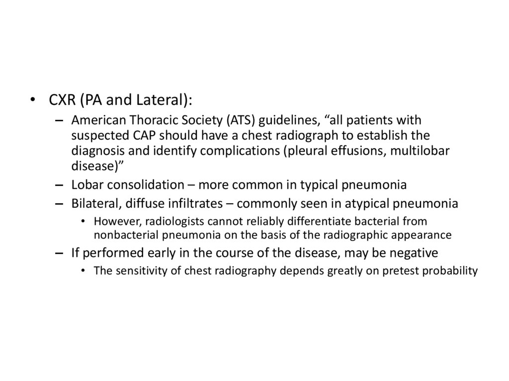
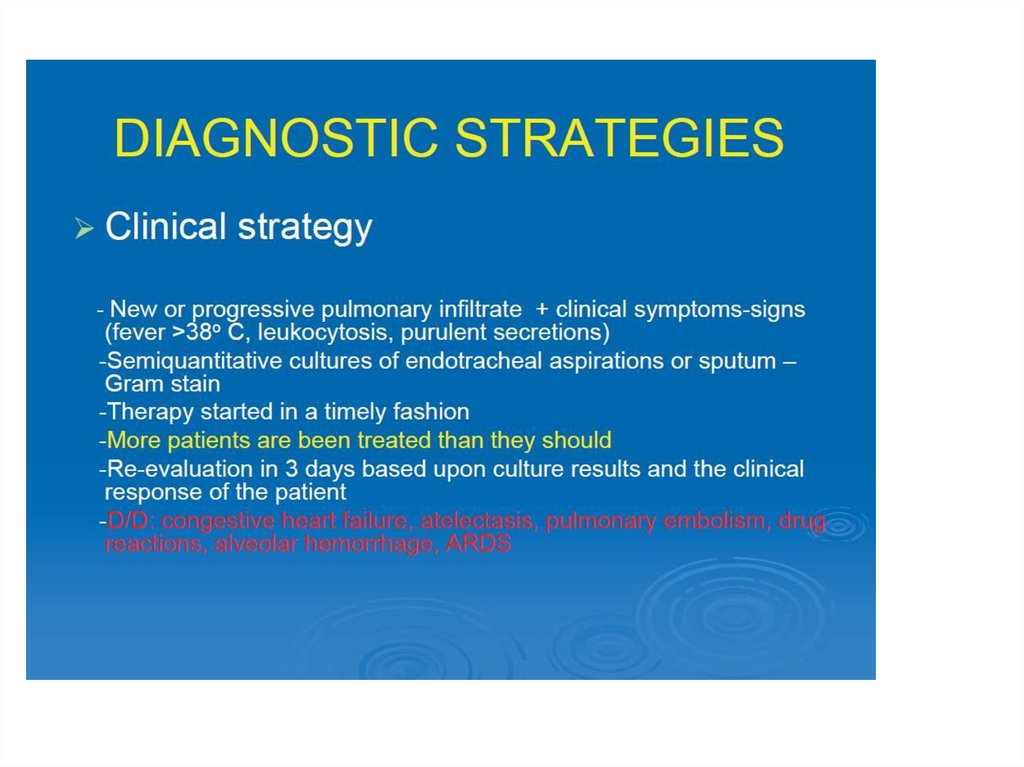
• CXR (PA and Lateral):– American Thoracic Society (ATS) guidelines, “all patients with
suspected CAP should have a chest radiograph to establish the
diagnosis and identify complications (pleural effusions, multilobar
disease)”
– Lobar consolidation – more common in typical pneumonia
– Bilateral, diffuse infiltrates – commonly seen in atypical pneumonia
• However, radiologists cannot reliably differentiate bacterial from
nonbacterial pneumonia on the basis of the radiographic appearance
– If performed early in the course of the disease, may be negative
• The sensitivity of chest radiography depends greatly on pretest probability
15.
16.
17.
18.
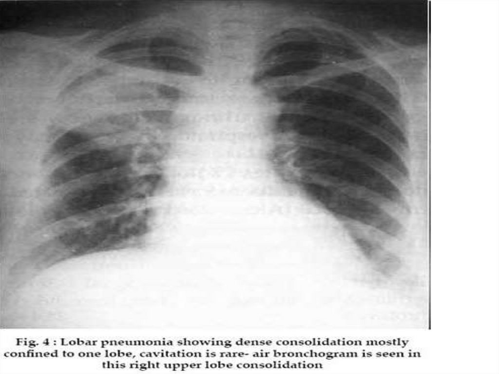
Lobar pneumonia• (also known as a non-segmental
pneumonia or focal non-segmental
pneumonia 7) is a radiological pattern associated
with homogenous,
fibrinosupparative consolidation of one or more
lobes of a lung in response to a bacterial
pneumonia.
• Streptococcus pneumoniae is the most common
causative organism of lobar pneumonia.
19.
Other causative organismsKlebsiella pneumoniae
Legionella pneumophila
Haemophilus influenzae
Mycobacterium tuberculosis
20.
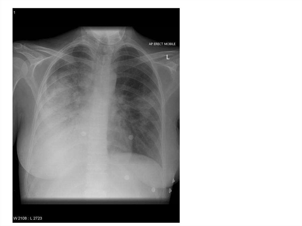
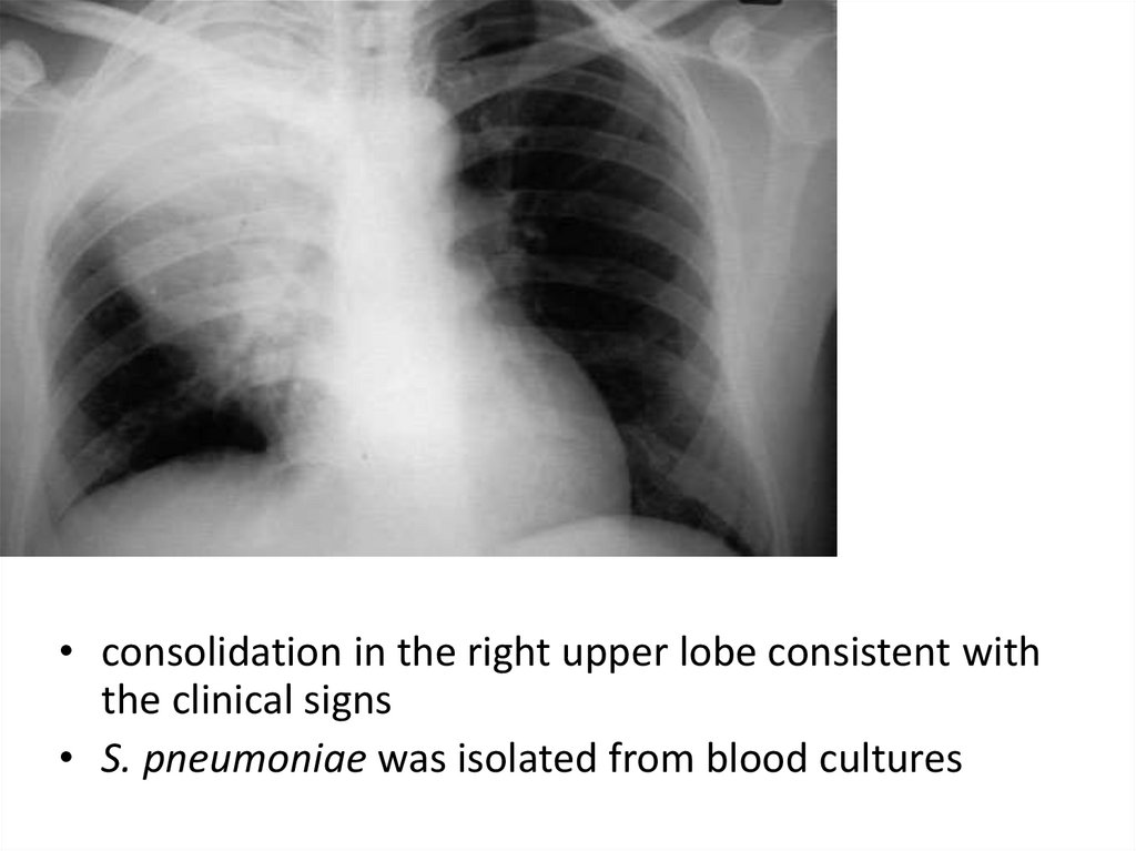
• consolidation in the right upper lobe consistent withthe clinical signs
• S. pneumoniae was isolated from blood cultures
21.
22.
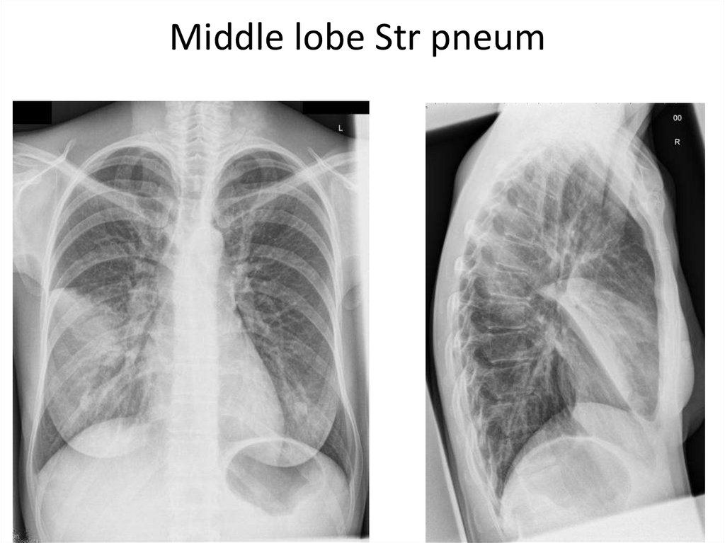
Middle lobe Str pneum23.
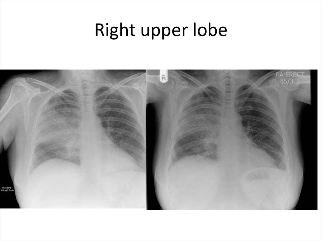
Right upper lobe24.
• consolidation in the right upper lobe consistent withthe clinical signs
• S. pneumoniae was isolated from blood cultures
25.
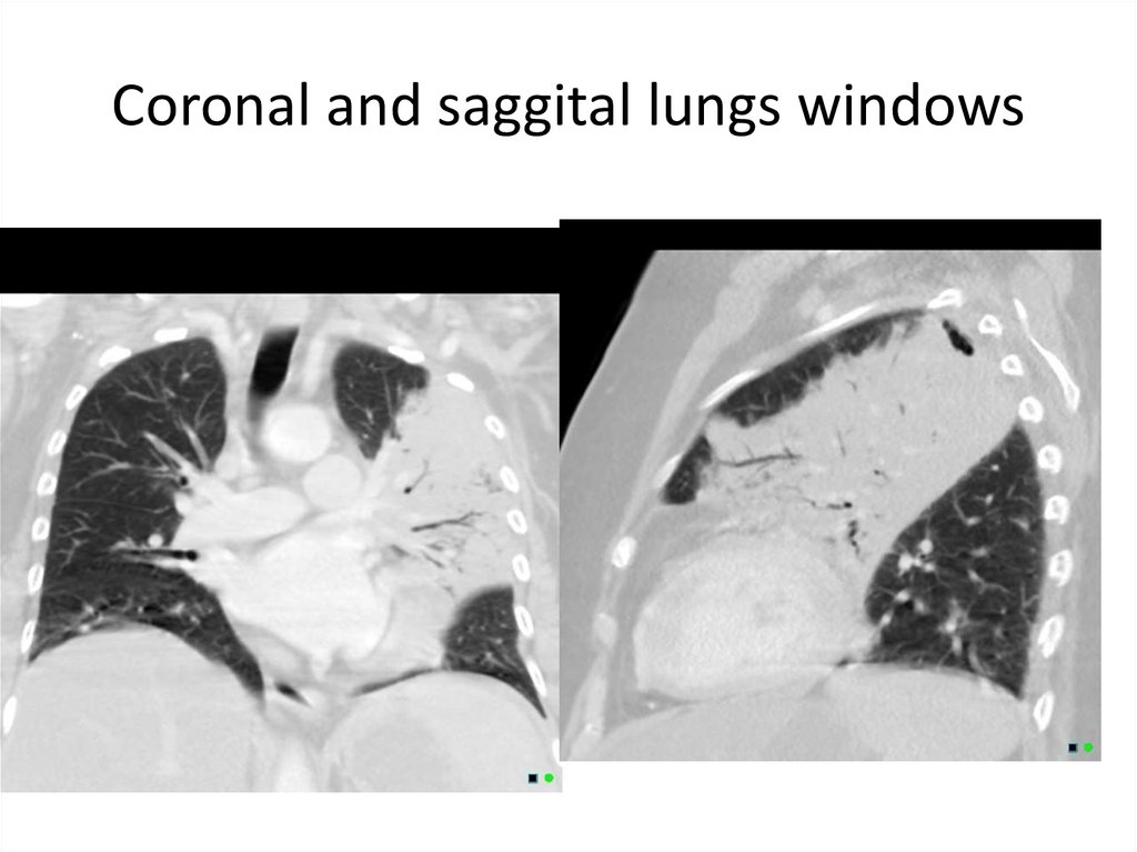
Coronal and saggital lungs windows26.
27.
28.
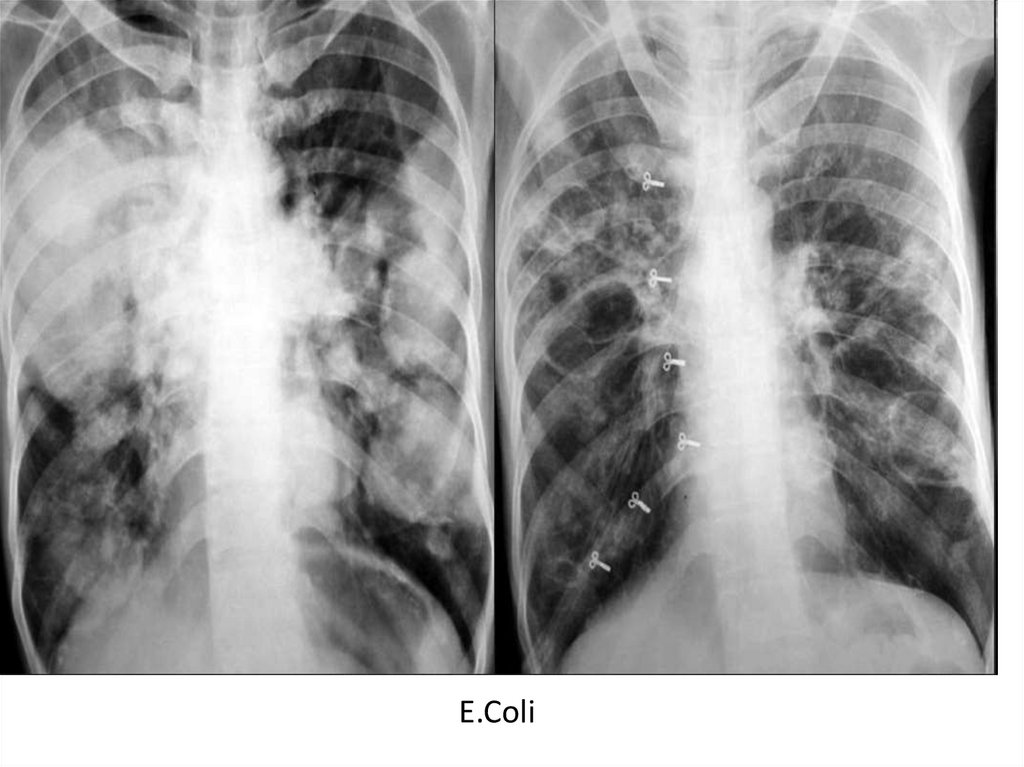
E.Coli29.
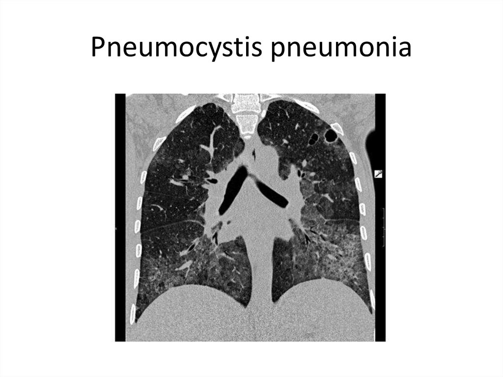
Pneumocystis pneumonia30.
31.
E.Coli32.
33.
34.
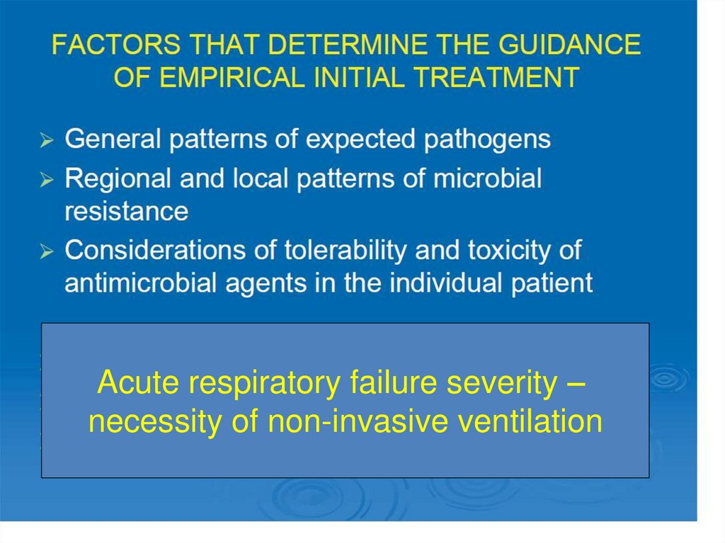
Acute respiratory failure severity –necessity of non-invasive ventilation
35.
36.
37.
38.
39.
40.
41.
42.
43.
44.
45.
46.
47.
48.
49.
50.
51.
52.
53.
54.
55.
56.
57.
58.
59.
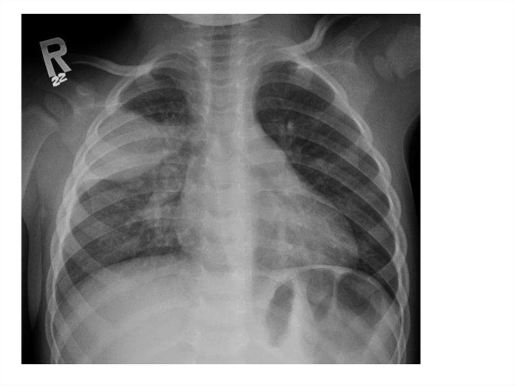
• Round pneumonia: usually seen in paediatricpatients. They are well defined, rounded
opacities that represent regions of infected
consolidation.
• Epidemiology
• mean age - 5 years and 90% of patients who
present with round pneumonia are younger than
twelve 5.
• uncommon after the age of eight because
collateral airways tend to be well developed by
this age 2,5.
60.
61.
62.
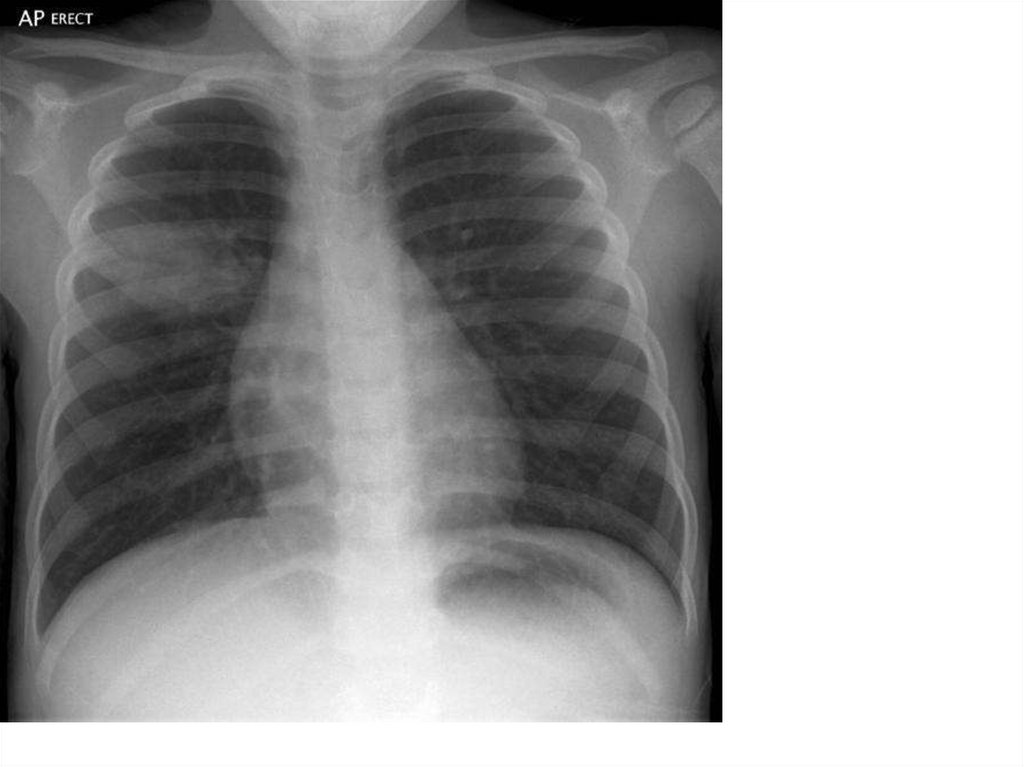
Bronchopneumonia• also sometimes known as lobular pneumonia
• radiological pattern associated with suppurative
peribronchiolar inflammation and subsequent
patchy consolidation of one or more secondary lobules
of a lung in response to a bacterial pneumonia.
• radiological appearance of bronchopneumonia is not
specific to any single causative organism, although
there are organisms which classically have a
radiological presentation of bronchopneumonia and
hence the identification of bronchopneumonia can
provide information regarding the likely aetiological
pathogens
63.
Causative organisms of a bronchopneumonia pattern include 3:
Staphylococcus aureus
Klebsiella pneumoniae
Haemophilus influenzae
Pseudomonas aeruginosa
Escherichia coli
Anaerobes, such as Proteus species
Histologically, multiple small foci of inflammation can be
demonstrated. Extensive congestion and dilation of bloods vessels
and areas of poorly circumscribed consolidation can be seen in
affected areas 8. These areas of inflammation are seperated by
areas of normal lung parenchyma 3.
64.
Radiology• Plain film
• Bronchopneumonia is characterised by multiple small nodular or
reticulonodular opacities which tend to be patchy and/or confluent.
This represents areas of lung where there are patches of
inflammation separated by normal lung parenchyma. 2.
• The distribution is often bilateral and asymmetric, and
predominantly involves the lung bases 8.
• CT - HRCT chest
• Multiple foci of opacity can be seen in a lobular pattern, centred at
centrilobular bronchioles. This may result in a tree-in-bud
appearance. These foci of consolidation can overlap to create a
larger heterogeneous confluent area of consolidation or 'patchwork
quilt' appearance 6.
65.
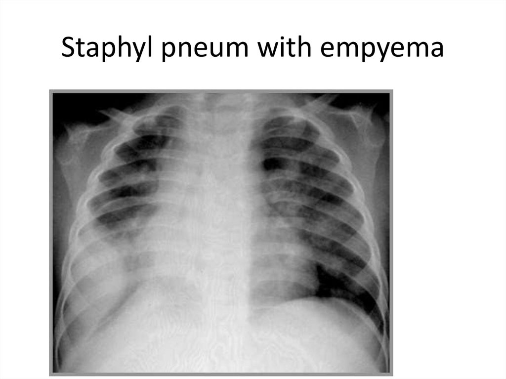
Staphyl pneum with empyema66.
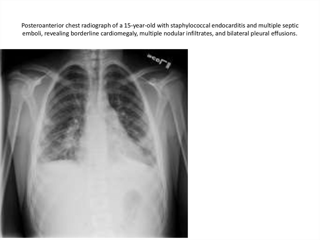
Posteroanterior chest radiograph of a 15-year-old with staphylococcal endocarditis and multiple septicemboli, revealing borderline cardiomegaly, multiple nodular infiltrates, and bilateral pleural effusions.
67.
Lat/view68.
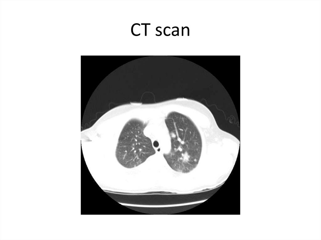
CT scan69.
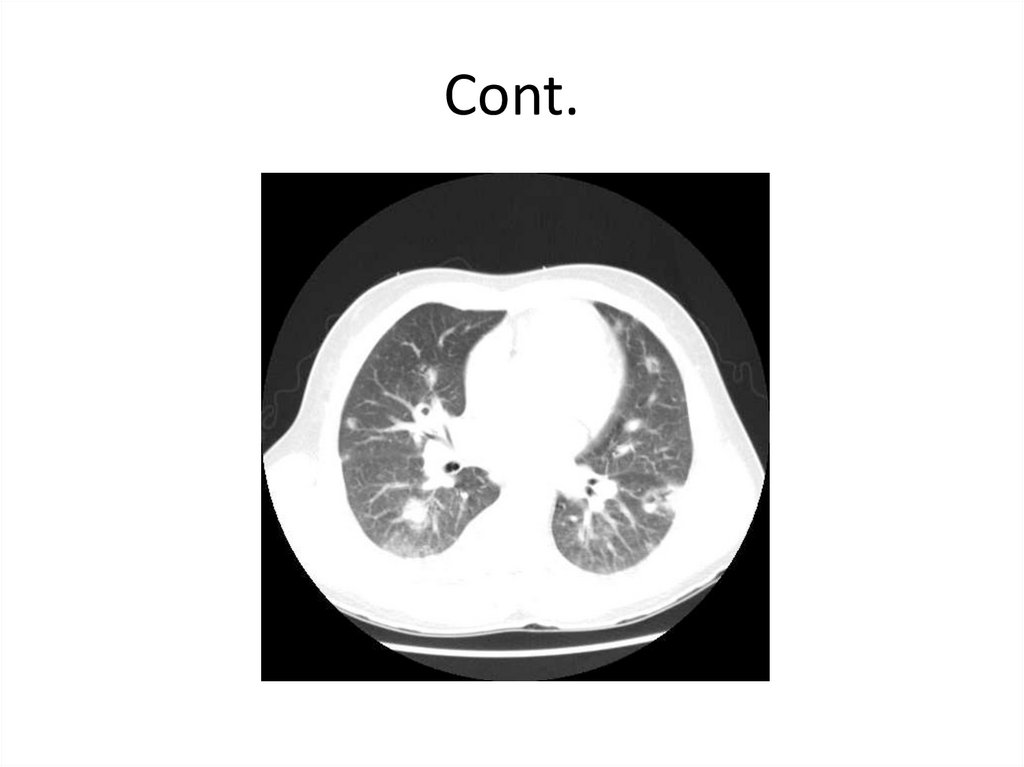
Cont.70.
same71.
72.
T scan of the thorax (mediastinalwindows) .
73.
follow up74.
75.
Cavitating clebsiella pneum76.
Mycoplasma77.
78.
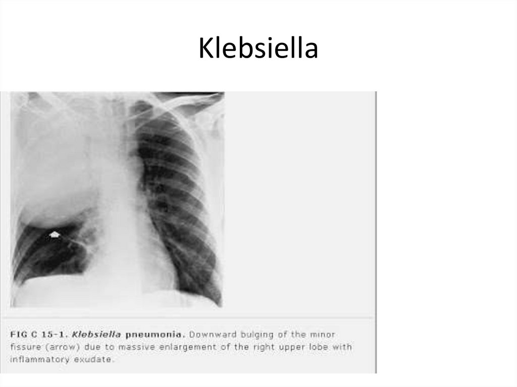
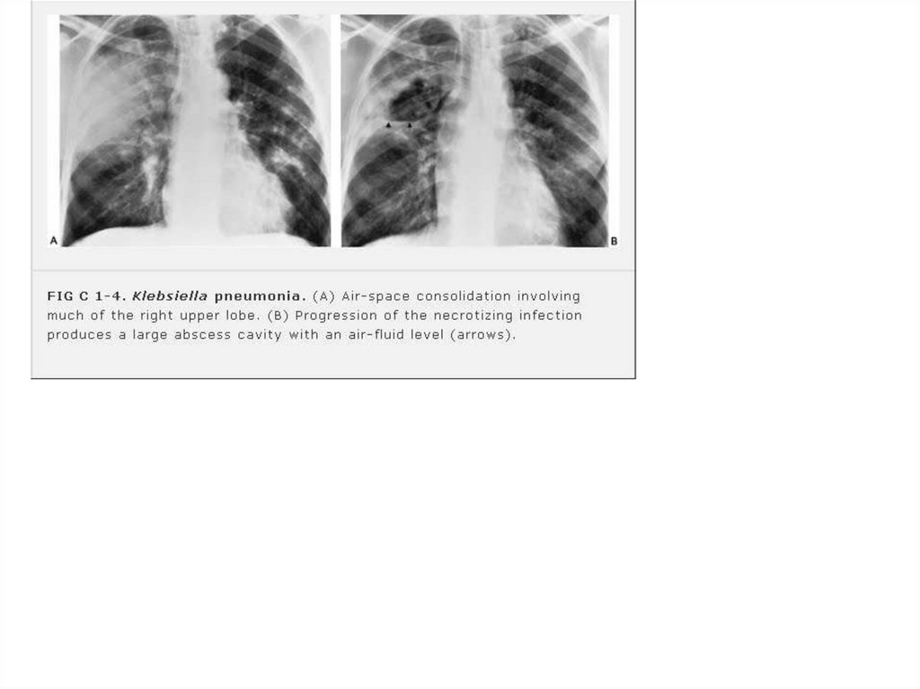
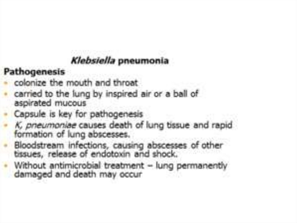
Klebsiella79.
80.
81.
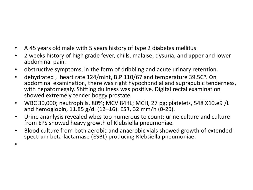
A 45 years old male with 5 years history of type 2 diabetes mellitus
2 weeks history of high grade fever, chills, malaise, dysuria, and upper and lower
abdominal pain.
obstructive symptoms, in the form of dribbling and acute urinary retention.
dehydrated , heart rate 124/mint, B.P 110/67 and temperature 39.5Co. On
abdominal examination, there was right hypochondial and suprapubic tenderness,
with hepatomegaly. Shifting dullness was positive. Digital rectal examination
showed extremely tender boggy prostate.
WBC 30,000; neutrophils, 80%; MCV 84 fL; MCH, 27 pg; platelets, 548 X10.e9 /L
and hemoglobin, 11.85 g/dl (12–16). ESR, 32 mm/h (0-20).
Urine ananlysis revealed wbcs too numerous to count; urine culture and culture
from EPS showed heavy growth of Klebsiella pneumoniae.
Blood culture from both aerobic and anaerobic vials showed growth of extendedspectrum beta-lactamase (ESBL) producing Klebsiella pneumoniae.
82.
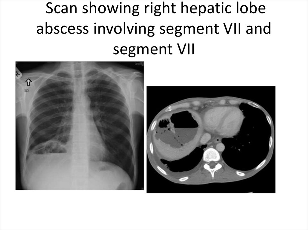
Scan showing right hepatic lobeabscess involving segment VII and
segment VII
83.
84.
85.
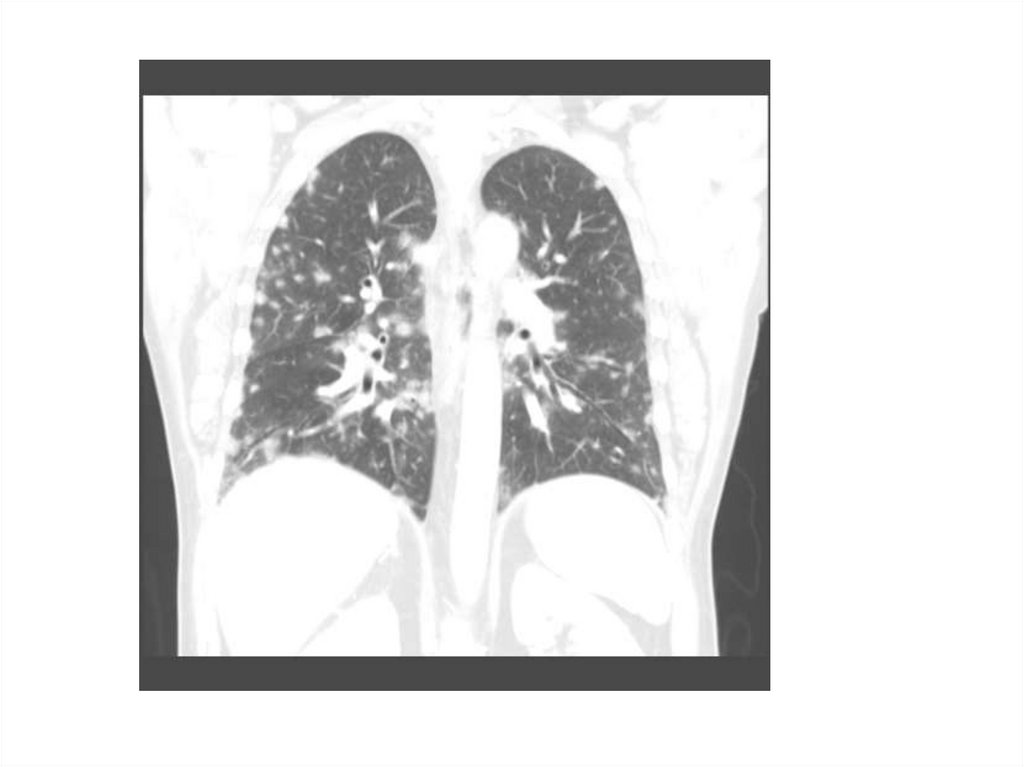
• The CT demonstrates multifocal opacities with somecavitation on the larger lesions. There seems to be a
peripheral and lower-lobe predominence. This could
represent atypical pneumonia (legionella, mycoplasma,
chlamydia), fungal pneumonia (cocciodomycosis,
histoplasmosis, aspergillosis), miliary tuberculosis,
metastatic lesions or carcinomatosis, septic emboli, or
viral pneumonia.
• After a significant inpatient workup the final diagnosis
was Human Metapneumovirus. All others were ruled
out and viral testing revealed this culprit.
86.
87.
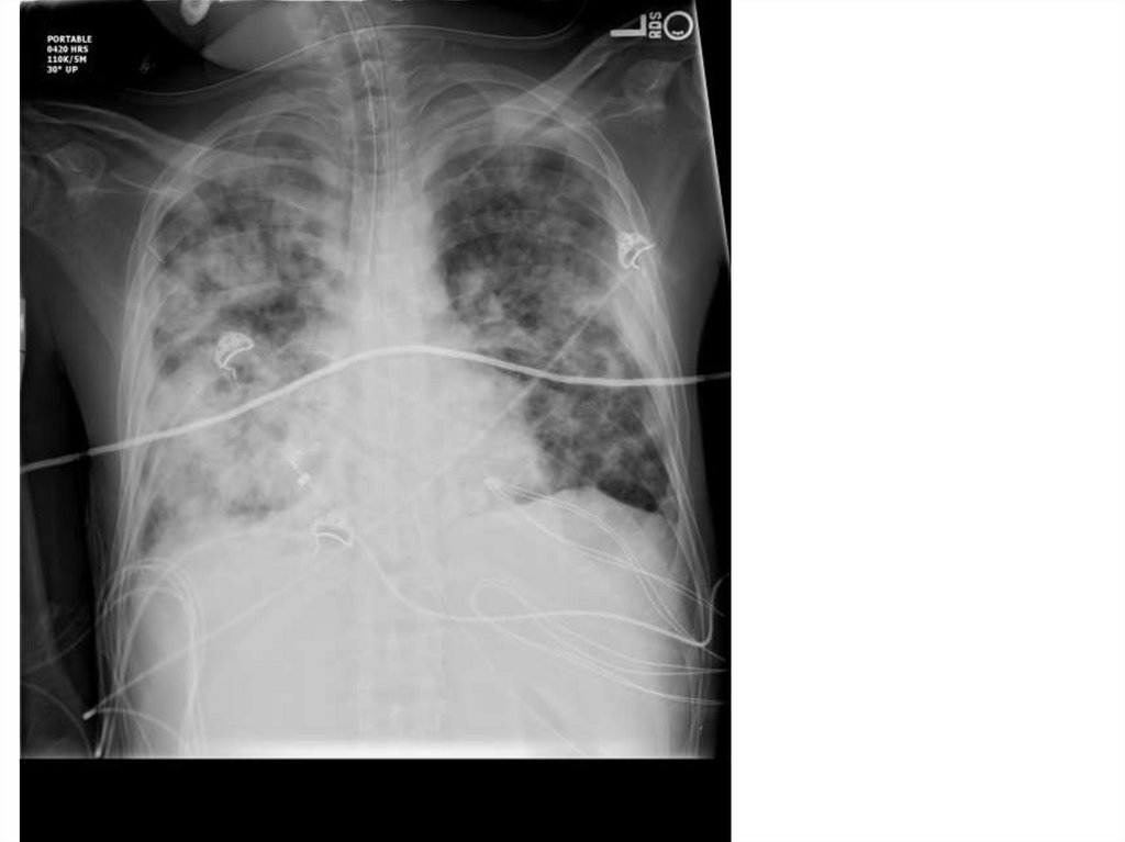
• This is a multilobar pneumonia vs. ARDS (Acute RespiratoryDistress Syndrome). AIDS patients can have the same
bacterial causes of multilobar pneumonia that is present in
other patient populations (Streptococcus pneumoniae,
Staphylococcus aureus, Haemophilus Influenza, Moraxella
catarrhalis, Mycoplasma pneumoniae, Chlamydophila
pneumoniae, Legionella pneumoniae, etc.). If they are
healthcare associated or hospital-acquired further drugresistant bugs such asPseudomonas aeruginosa and MRSA
could be implicated. Infectious organisms specifically
involved in immunocompromised hosts could include
(among others):
88.
Multilobar infiltratesPneumocystis Jiroveci (PCP pneumonia)
Coccidioides species
Cytomegalovirus (CMV)
Tuberculosis (TB)
Histoplasma species
Aspergillus species
Mycobacterium avium complex (MAC)
Influenza
Herpes simplex virus (HSV)
Varicella-zoster virus (VZV)
Legionella species
Nocardia species
Cryptococcus neoformans
Mucoraceae species
Strongyloides species
Toxoplasma species
Capnocytophaga species
89.
Non-infectious causes ofmultilobar infiltrates
diffuse alveolar hemorrhage,
cardiogenic pulmonary edema,
ARDS,
multilobar involvement of the Xray above could
implicate certain pathogens in favor of others (for
example, Pneumocystis Jiroveci is usually multilobar as
opposed to Streptococcus pneumonia which usually
will cause a dense, lobar pneumonia).
• CMV rather than a bat-wing ground-glass appearance
ofPneumocystis Jiroveci. For further discussion on
pneumonia radiographic findings in AIDS, please
see radiopaedia.org discussion below:
90.
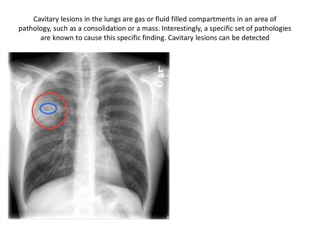
Cavitary lesions in the lungs are gas or fluid filled compartments in an area ofpathology, such as a consolidation or a mass. Interestingly, a specific set of pathologies
are known to cause this specific finding. Cavitary lesions can be detected