


")


")


")


 Медицина
Похожие презентации:


Pneumonia in children. Diagnostics and treatment
1. Pneumonia in children. Diagnostics and treatment.
12. Plan of the lecture
Definition pneumonia2. Etiology
3. Pneumonia pathogenesis
4. Classification of
pneumonia
5. Pneumonia treatment
1.
2
3. Pneumonia is a group of acute focal infectious inflammatory diseases varied in etiology, pathogenesis and morphologic characteristic with predominant involvement in pathologic process of respiratory tract with invariable presence of alveolar inflammatory
Pneumonia is a group of acute focalinfectious inflammatory diseases
varied in etiology, pathogenesis and
morphologic characteristic with
predominant involvement in pathologic
process of respiratory tract with
invariable presence of alveolar
inflammatory exudate.
3
4. Predisposed anatomy-physiologic peculiarities in children to pneumonia
Trachea and big bronchi are short and wide – easy penetrationof infection
Little bronchi and bronchioli are narrow and are deficient in
connective and muscular tissue – they are easily collapsed and
obstructed
Inadequate drainage of several segments due to peculiarities of
bronchial branching – frequent involvement of I, II, IX, X, VI
segments bilateral and of IV, V segments of left lung
Lack of elastic fibers and surfactant –lung rigidity, inclination
to atelectasis and emphysema development
Insufficient mucocilliar clearance – difficulties in foreign bodies
removing
Insufficient synthesis of interferon and IgA – incompatibility
immune response
Plethoric lung parenchima, rich in interstitial vascularization; in
perinatal period is collapsed
4
5. Predisposing premorbid factors for pneumonia
Premature newbornsSevere perinatal pathology: prenatal hypoxia, asphyxia, intrapartum
trauma
Vomiting and regurgitation syndrome
Artificial feeding
Constitution anomalies
Rickets
Malnutrition
Congenital heart diseases
Cystic fibrosis
Congenital lung malformations
Surgical treatment
Inherited immunodeficiencies
Hypovitaminosis
Chronic focuses of infection
Smoking
5
6. Pneumonia etiology
Streptococcus Pneumonia ( 60-80% cases ofcommunity acquired pneumonia
Hemophilus influenzae
Moraxella Catarrhalis
In newborns and infants – Staphylococcus,
gram (-) microflora
Mycoplasma pneumonia, Chlamidia psittaci,
Chl.pneumonia (10-12%).
Severe pneumonia are caused by mixed micriflora
Pneumocystis pneumonia can develop only in immune
compromised host (deep prematurity, combined
immunodefficiancy, AIDS, imunosuppression)
Viral pneumonia is rare disease. It can be caused
by flu, (hemorrhagic pneumonia,), in bronchiolitis,
adenoviral and RS viral infection
6
7. All microorganisms from sputum are divided into 3 groups
pathogenicprovisional pathogenic
nonpathogenic
7
8. Pathogenic are microorganisms with complementary receptors to surface cell receptors in respiratory tract. It gives them opportunity to adhere and multiply on mucus membrane of respiratory tract. They are Pneumococcus, Hemophylus influenza, Legionella, My
Pathogenic are microorganisms with complementary receptorsto surface cell receptors in respiratory tract. It gives them
opportunity to adhere and multiply on mucus membrane of
respiratory tract.
They are Pneumococcus, Hemophylus influenza, Legionella,
Mycoplasma, Ricketsia, Mycobacterium tuberculosis etc.
Provisional pathogenic are microorganisms that have no
receptors and can’t be fixed on epithelium. Protective
mechanisms can easily eliminate them. Only impairment of
these mechanisms lead for their penetration, spreading and
multiplying ( ARD, overcooling, immune suppression etc)
Nonpathogenic microbes –microorganisms that can cause
inflammation only in cases of severe degree of
immunodeficiency. They are aerobe and anaerobe
saprophytes from upper respiratory tract.
8
9. Diagnostic criteria of bacterial pneumonia
Anamnestic dataHospital acquired pneumonia is developed in 48 hours after hospitalization
and 48 h after discharging from hospital
Bacterial intoxication symptoms
Clinical:
Fever more than 3 days
Tachycardia
Paleness, regurgitation
Lab data:
Neutrophyl leukocytosis
Elevated ESR
Functional respiratory disturbancies
Increased respiratory rate more than 20% from age norma
Accessory musculature involving in respiration
Cough or its equivalents
Cyanosis ( perioral, periorbital, diffuse)
Local symptoms in pneumonia:
Percussion sound shortening ( dullness)
Breathing sound conductivity changes (attenuation, rales)
Radiologic confirmation
9
10. Pneumonia classification in children
Clinicalform
Contamination
Course
Focal
Community
aquired
(home)
Acute
Segmenta Hospital or
l
Nosocomial
Due to
Focal
perinatal
Confluent Infection
Croupous
Interstiti
al
In patients
with
immune
deficiency
(less than
6 weeks)
Lingerin
g
( more
than 6
weeks to
8 mo)
Recurre
nt
Complications
Pulmonic
Synpneum
onial
pleuritis
Methapne
umonial
pleurisy
Pulmonary
destruction
Lung
abscess
Extrapulmonic
Infectioustoxic shock
DIC-syndrome
Cardiovascular
insufficiency
Respiratory
distress
Syndrome
Toxic affection
of
Pneumotho other organs
( carditis,
rax
nephritis,
hepatitis,
Pyopneumo acute kidney
failure, otitis,
osteomyelitis
10
thorax
etc)
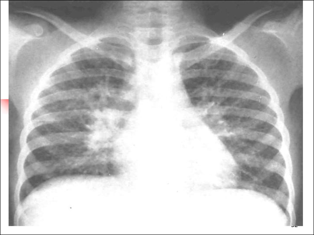
11. Focal pneumonia (30-40% of pneumonia)
It frequently starts from bronchi –bronchopneumonia
Frequently developed after ARD
Cough is deep and moist
Intoxication
Respiratory failure can be present
Percussion pulmonary clear sound or even with
resonance sound but under the focus shortening of
the sound
Auscultation: focal bubbling rales, focal crepitation
If accompanied by bronchitis – bilateral dry and
moist rales
Radiologic picture presence of interstitial
11
involvement with focal infiltration of 1,5 cm in
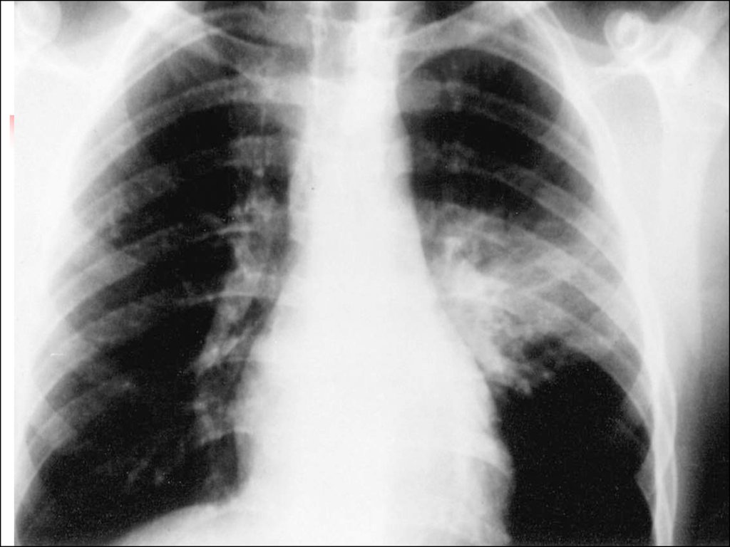
12. Focal-confluent pneumonia
Several segments are affected or the whole lobewith focal pulmonary destruction. Intoxication
is prominent, massive lung tissue involvement,
usually pleurisy.
As a rule ARD precedes with progressive course
with involvement of bronchi.
Radiologic peculiarities
Infiltrative shadows are not homogeneous
Process usually is unilateral more frequently in
right lung
At affected side intercostal and lobe pleura
reaction is present
12
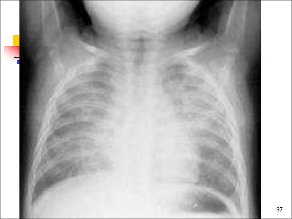
13. Segmental Pneumonia
Pneumonia affects one or several segments. Moist rales are nottypical or they disappear very quickly.
There are 3 types of course:
With good prognosis, without symptoms
Course is like in croupous pneumonia – sudden onset with
fever and cyclic course. Pains in abdomen and chest
Clinical picture like in focal pneumonia, but auscultative data
are vague, percussion isn’t clear. Frequent pleuricy, atelectasis
Inclination for abscess formation, destruction, lingering course
X-ray signs: more frequent localization in 1,3 segments of
right and 8, 9, 10 segments of both lungs, in 5,4 segments of
left lung
Process is unilateral as a rule
Regional lymph nodes are increased on affected side
Pleural ( costal or interlobular) reaction is visible
Duration of pneumonia 10-12 days
13
More frequent complications : atelectasis, pleuritis, destruction
14. Interstitial pneumonia (1% of all pneumonia)
Acute inflammation of interstitium and less manifestedaffection of broncho alveolar structures
Paleness is typical
Pertussis –like cough
Tympanic resonance during percussion
Respiratory sound is rough, irregular dry and various
moist bubbling rales
Prominent respiratory failure
Pathogen can’t be revealed in common way
More frequent causative factors are fungus,
Pneumocystis, Chlamidia, Mycoplasma,
14
Ricketsia, Legionellas
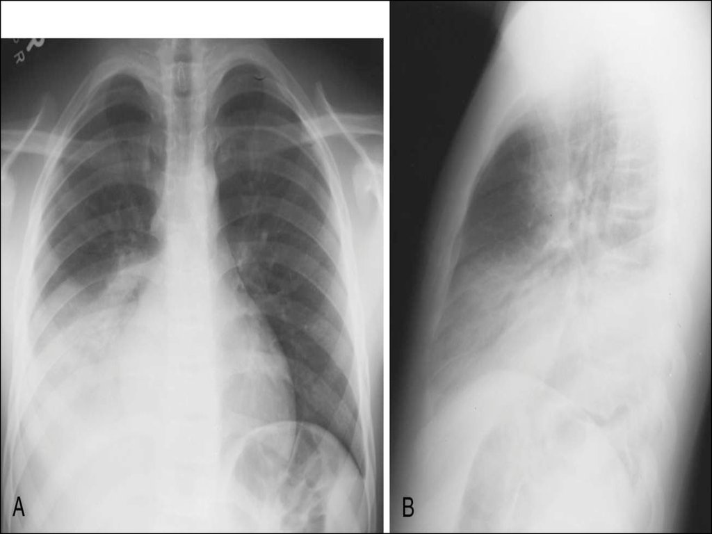
15. Croupous pneumonia
Classic example of community acquired pneumonia. It islobe
or segment affection with pleura involvement
(pleuropneumonia).
It’s difficult to differ it from segmental pneumonia only
radiologically. Clinical picture plays the clue role
Acute onset
Cyclic course
Febrile or high febrile fever, flush red on affected side
Sputum is rusty, herpes labialis and nasalis
Lung destruction is very rare
Localization in lower lobes
Chest pain due to pleuritis
Abdomen pain like in appendicitis
15
Meningeal form of pneumonia
16. Respiratory Failure –is a condition of disturbed gaseous blood composition due to lung function failure or when maintaining of proper partial O2 and CO2 containing is achieved by forcing of external respiratory structures that produce functional exhaust
Respiratory Failure –is acondition of disturbed gaseous
blood composition due to lung
function failure or when
maintaining of proper partial O2
and CO2 containing is achieved
by forcing of external
respiratory structures that
produce functional exhaustion
of organism.
16
17. Clinical classification of respiratory failure
Grade IDyspnea after loading, in rest dyspnea is absent. Accessory
musculature
isn’t involved, irregular perioral cyanosis more visible after
agitation. BP is
normal. HR ratio to RR=3,5-2,5 : 1`, tachycardia. Blood gases
composition: PaCO2 <4,67 Kpa : Pa O2=8,76-10 kPa
Grade II
Dyspnea in rest, accessory musculature involvement, retractions
in chest,
constant acrocyanosis, BP is elevated, tachycardia, flaccidity,
drowsiness,
adynamia. HR ratio RR = 2-1,5 : 1: PaO2= 7,33-8,53 kPa: PaCO2
= 4,67-5,87 kPa
Grade III
Manifested dyspnea ( more than 50% from N). Bradypnoe and
17
dyspnoe,
18. Main principles of pneumonia treatment
Treatment must be opportuneand integrated
Etiotropic therapy directed for
eradication of pathogen
Treatment of pathologic
syndromes, complications and
co-morbidities
Rational rehabilitation process
18
19. Indications for hospitalization
InfantsRespiratory failure, necessity of oxygen
therapy, manifested intoxication
Dehydration, impossibility of oral
drinking
Unfavourable premorbid condition,
immune deficiency, developmental
anomalies
Suspicion as for Staphylococcal etiology,
complications like pleuritis. Ineffective
home treatment within 24-36 hours
19
20. Pay attention for
Respiratory rate ( mainindex). In children 2-12 mo
old
RR> 50/min and
for children
12 mo- 5 y.o
RR>40/min is threatening.
Retractions of chest lower
part
Stridor
20
21. It’s important
Air humidification in room where child ispresent
Clothes must be suitable, surrounding
temperature must be optimal
Main task is normalization of nose passage
of air
Sleeping must be organized with raised
head part of bed
Parents mustn’t prohibit child to cough
To provide with proper intake of liquids
intake by oral or parenteral way
21
Feeding must be usual for age enriched by
22. Etiotropic therapy
Foundation of etiotropictreatment is empiric start
antibiotic therapy with following
its correction
Empiric start antibacterial
therapy is performed depending
on expected causative factor
22
23. Main groups of antimicrobial drugs
Main groups of antimicrobialBeta-lactams
drugs
1. Penicillines
2. Cephalosporines
3. Monobactams (Aztreonam)
4. Carbapenems (Imipenem, Meropenem)
Aminoglycosides
Fluoroquinolones
Macrolides
Glycopeptides
Nitromidazolines
Tetracyclines
Chloramphenicol
Lyncosamines
Nitrophuranes
Sulfanilamides
Antituberculosis
Antifungal
23
24. Main statements of antibiotic therapy
Antibiotic administration must peroral incommunity acquired uncomplicated pneumonia
In case of severe course only parenteral antibiotic
administration, combinations of antibiotics
Ineffectiveness of beta-lactams indicate resistant
or atypical microorganisms presence
Duration of uncomplicated community acquired
pneumonia is 7-10 days. In case of complications
duration must be not less than 14 days
In case of parenteral antibiotic administration
condition improvement demand change antibiotic
administration for oral intake so called step
approach
First antibiotic course mustn’t combined with
24
25. Efficacy criteria of antibiotic therapy in pneumonia
Efficacy assessment is performed inuncomplicated pneumonia 24-48 hours
after treatment beginning. If there are
some complications it is performed 4872 hours later
Main criteria:
Dynamics of common child’s condition
Disappearing of fever
Normalization of respiratory rate and Ps
and their ratio
25
Improving of lab and X-ray data
26. Effects of antibiotic therapy
Complete effect- temperature decreasing lessthan 38C 24-48 hours later in uncomplicated
pneumonia form or 72 hours later in complicated
pneumonia, improving of condition, appetite,
dyspnea reducing
Partly improving- temperature is higher 38C
with toxicosis resolving, appetite improving,
absence of negative radiologic dynamics
Effect absence – Constant high temperature
more than 38 C, condition worsening and/or
progressive worsening of lung and pleura
changes
26
27. Side effects of antibiotic medication
Allergic reactionsNephrotoxicity
Ototoxicity
Disbiosis
All antibiotics, predominantly
penicillines
Aminoglycosides, cephalosporines
Aminoglycosides
Cephalosporines, penicillines,
macrolides
Pseudomembranoes
colitis
Hepatotoxicity
Penicillines, cephalosporines
Cholestasis
Macrolides
Leucopoesis
supression
Chloramphenicol
Osteogenesis
disturbancies
Tetracyclines,lincomycin
Tetracyclines, cephalosporines
27
28. Pathogenic treatment
Respiratory supplementationaccording to respiratory failure
Desintoxication. If indications are
present intravenous infusion is
performed to correct acidic – basic
condition, fluid and electrolyte
disorders
Symptomatic treatment can
include antipyretics etc.
28
29. Segmental structure of lungs (scheme)
2930. Questions
To indicate etiologic and pathophysiologic factors atpneumonia in children
To classify pneumonia, respiratory failure, analyze typical
clinic of the pneumonia, respiratory failure in children.
To indicate aspects of the pneumonia in newborns and to
mace previous diagnose.
To make list of the examination and to analyze data of the
laboratory and instrumental examination.
To prescribe treatment, rehabilitation, prophylaxis of the
pneumonia in children.
To diagnose and to give the first medical aim in acute
respyratory failure in children.
To perform differential diagnostic of pneumonias in children
To make prognosis at pneumonia.
To demonstrate morally-deontological principles of the
30
subordination in the pulmonologic department
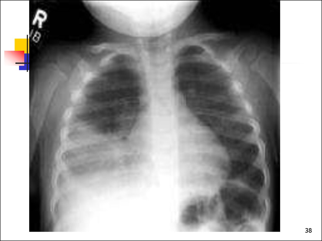
31. Pneumonia complication- pneumothorax
Pneumonia complicationpneumothorax31
32.
3233.
3334.
3435.
3536.
3637.
3738.
3839.
Thank you39