


 Медицина
Похожие презентации:
")


")


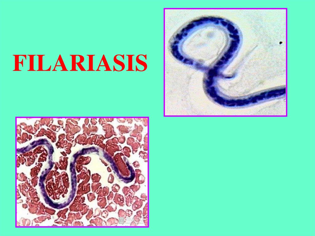
Filariasis
1.
FILARIASIS2.
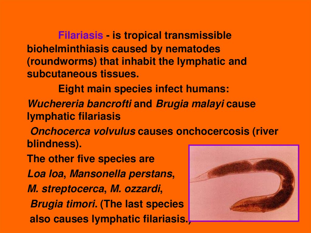
Filariasis - is tropical transmissiblebiohelminthiasis caused by nematodes
(roundworms) that inhabit the lymphatic and
subcutaneous tissues.
Eight main species infect humans:
Wuchereria bancrofti and Brugia malayi cause
lymphatic filariasis
Onchocerca volvulus causes onchocercosis (river
blindness).
The other five species are
Loa loa, Mansonella perstans,
M. streptocerca, M. ozzardi,
Brugia timori. (The last species
also causes lymphatic filariasis.)
3.
Lymphatic filariasis - Wuchereriasis andBrugiasis common in 76 countries, where the risk of
infection are susceptible to 905 million, of which 90
million are sick.
2\3 of affected live in China, India, Indonesia, many
countries of Africa and Pacific region;
4.
Onchocercosis - is distributed in 34 countries,mainly in tropical Africa, the Volta river basin,
Mexico, Columbia, Guatemala.
The number of patients 17.6 million, of which
26000 are blind.
5.
Loa-loa disease (loaosis) - is found only in theforest zone of West and Central Africa;
6.
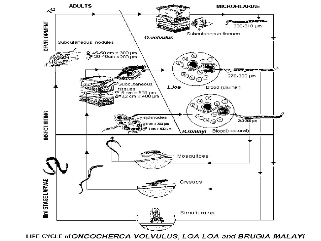
General properties of filariasis:1. They are – all biohelminths , developing with the change
of owners.
2. The final host – is human, intermediate hosts of – arthropods
(mosquitoes, midges, mokrets).
3. All filarias divide on male and female.
4. The adults (macrofilaria)dwell in various human tissues
where they can live for several years (subcutaneous tissue –
onchocercosis, loaosis, streptocercosis, lymph vessels –
wuchereriasis and brugiasis connective tissue etc).
5. Females produce a larvae (microfilaria) which penetrate into
blood stream or superficial skin layers (onchocercosis),
they do not grow and change morphologically.
6. Length of adult males up to 50 mm, females - up to 100 mm,
microfilaria - 0,3 mm.
7.
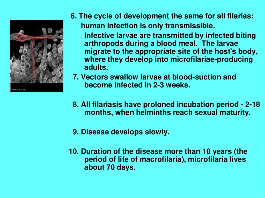
6. The cycle of development the same for all filarias:human infection is only transmissible.
Infective larvae are transmitted by infected biting
arthropods during a blood meal. The larvae
migrate to the appropriate site of the host's body,
where they develop into microfilariae-producing
adults.
7. Vectors swallow larvae at blood-suction and
become infected in 2-3 weeks.
8. All filariasis have proloned incubation period - 2-18
months, when helminths reach sexual maturity.
9. Disease develops slowly.
10. Duration of the disease more than 10 years (the
period of life of macrofilaria), microfilaria lives
about 70 days.
8.
9.
11. There are three groups of filariasis depending onthe concentration of larvae in the peripheral
blood:
- periodical –peak of the highest concentration of
larvae in peripheral blood is observed in a day or
at night, in other times filarias are absent ,
- subperiodical - larvae is constantly present in
the peripheral blood, but the highest
concentration may be seen only in the same time
of the day.
- non-recurrent – microfilarias are allways
present in the blood in constant concentration.
Frequency coincides with the period of
maximal activity of the vector.
10.
Wuchereriasis and Brugiasis(Filariasis Bancrofti, F. Malayi)
- helminthiasis affecting the lymphatic system.
ETIOLOGY: causative agent of
wuchereriasis – Wuchereria bancrofti
(Wucherer and Bancroft - scientists, who have described the
helminth),
brugiasis - Brugia malayi (Brug – scientist)
Macrofilarias parasites in the lymph nodes and vessels,
microfilarias - in the blood.
EPIDEMIOLOGY: source of infection
in W.(anthroponosis) – man,
in B. (zoonosis) – human, cats, dogs, monkeys.
11.
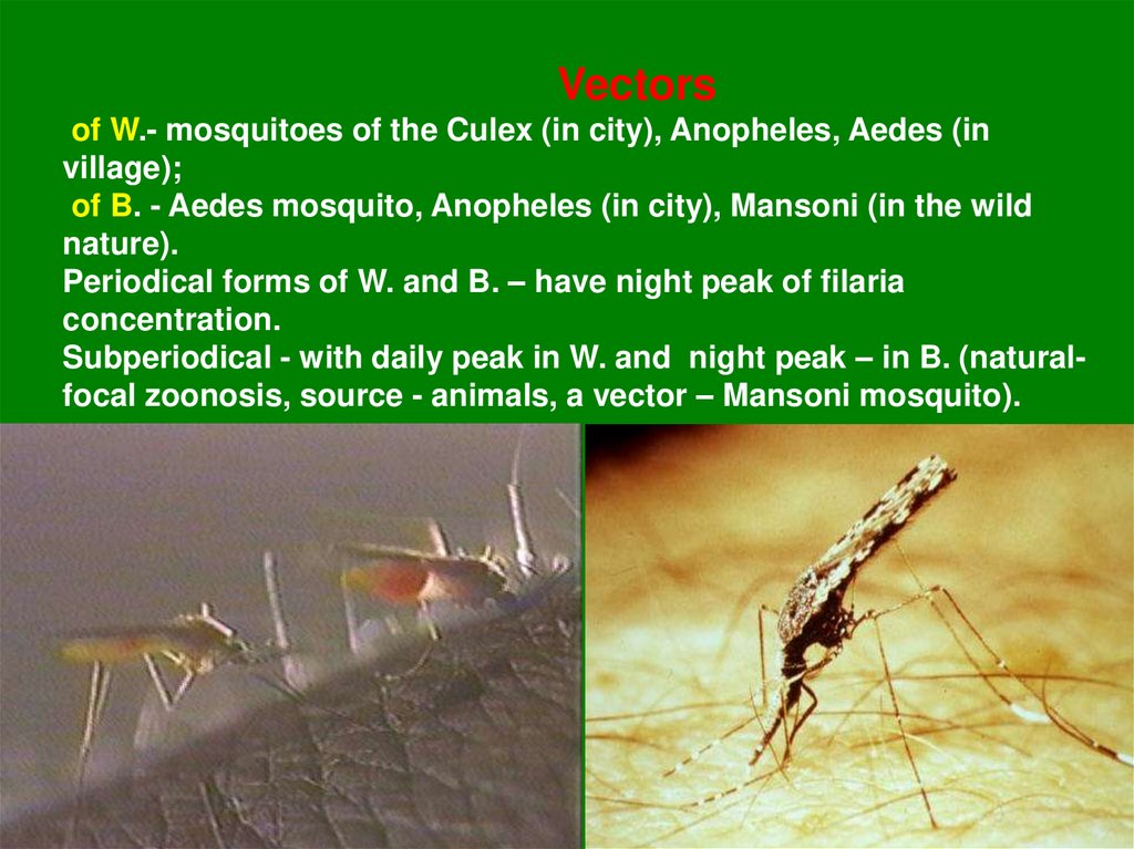
Vectorsof W.- mosquitoes of the Culex (in city), Anopheles, Aedes (in
village);
of B. - Aedes mosquito, Anopheles (in city), Mansoni (in the wild
nature).
Periodical forms of W. and B. – have night peak of filaria
concentration.
Subperiodical - with daily peak in W. and night peak – in B. (naturalfocal zoonosis, source - animals, a vector – Mansoni mosquito).
12.
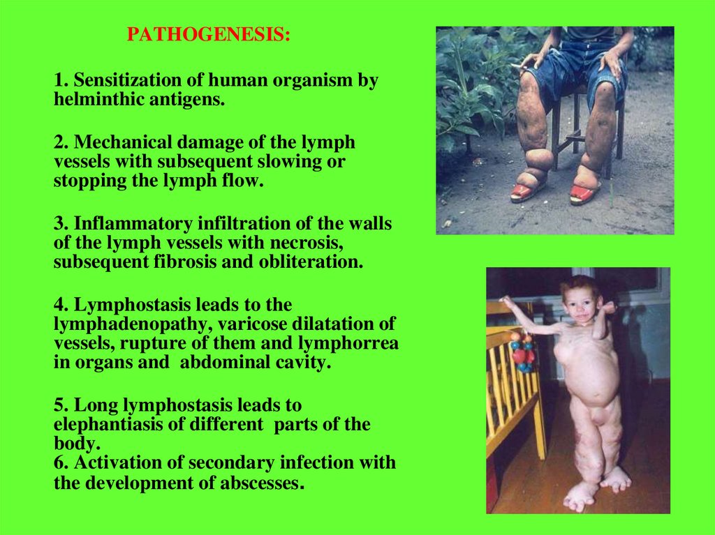
PATHOGENESIS:1. Sensitization of human organism by
helminthic antigens.
2. Mechanical damage of the lymph
vessels with subsequent slowing or
stopping the lymph flow.
3. Inflammatory infiltration of the walls
of the lymph vessels with necrosis,
subsequent fibrosis and obliteration.
4. Lymphostasis leads to the
lymphadenopathy, varicose dilatation of
vessels, rupture of them and lymphorrea
in organs and abdominal cavity.
5. Long lymphostasis leads to
elephantiasis of different parts of the
body.
6. Activation of secondary infection with
the development of abscesses.
13.
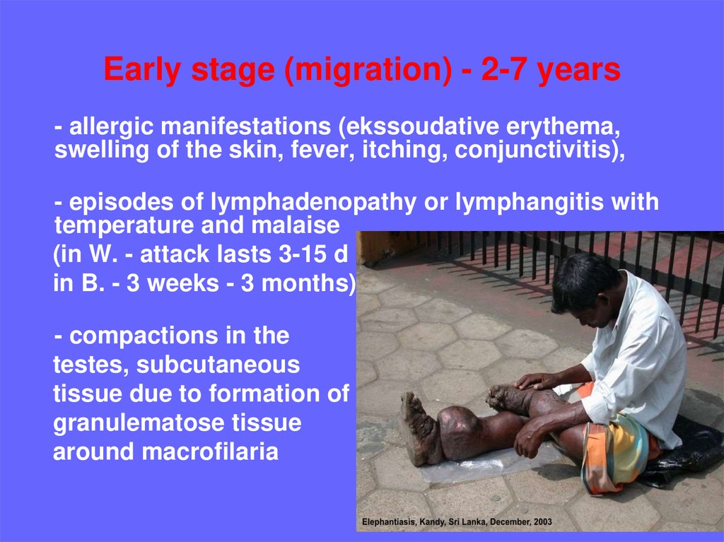
Early stage (migration) - 2-7 years- allergic manifestations (ekssoudative erythema,
swelling of the skin, fever, itching, conjunctivitis),
- episodes of lymphadenopathy or lymphangitis with
temperature and malaise
(in W. - attack lasts 3-15 d
in B. - 3 weeks - 3 months)
- compactions in the
testes, subcutaneous
tissue due to formation of
granulematose tissue
around macrofilaria
14.
- funikulit, epididymitis, orchitis (in W.)- abscesses in the upper parts of the thighs,
under the fascia of abdominal muscles.
They are sterile, appear and disappear
slowly (in W.)
-
- often the crotch lymphadenitis and
lymphangitis in inguinal area and axillar
region (seldom) – in B.
- eosinophilic pulmonary infiltrates,
hepatosplenomegaly, eosinophilia in CBC,
- often inflammation of lymph nodes and
abscesses (Indonesia, Malaysia, Thailand),
rarely - in India.
15.
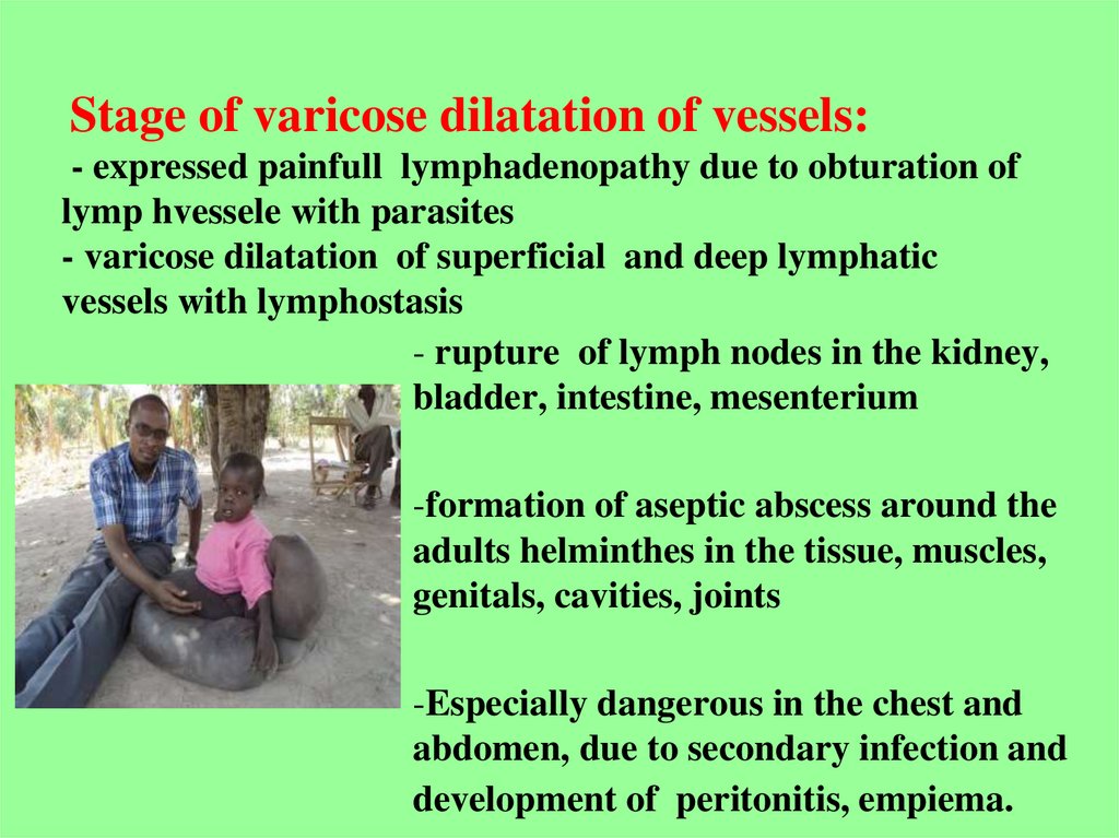
Stage of varicose dilatation of vessels:- expressed painfull lymphadenopathy due to obturation of
lymp hvessele with parasites
- varicose dilatation of superficial and deep lymphatic
vessels with lymphostasis
- rupture of lymph nodes in the kidney,
bladder, intestine, mesenterium
-formation of aseptic abscess around the
adults helminthes in the tissue, muscles,
genitals, cavities, joints
-Especially dangerous in the chest and
abdomen, due to secondary infection and
development of peritonitis, empiema.
16.
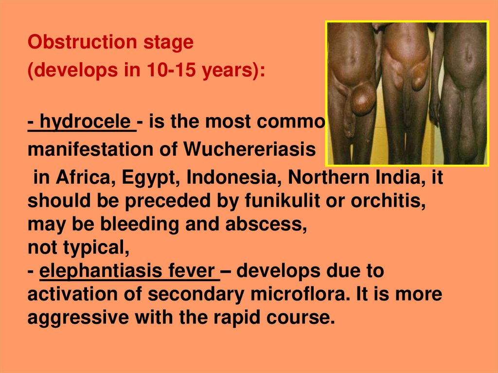
Obstruction stage(develops in 10-15 years):
- hydrocele - is the most common
manifestation of Wuchereriasis
in Africa, Egypt, Indonesia, Northern India, it
should be preceded by funikulit or orchitis,
may be bleeding and abscess,
not typical,
- elephantiasis fever – develops due to
activation of secondary microflora. It is more
aggressive with the rapid course.
17.
18.
-Swelling spreads - the foot, ankle, thigh-extremity increases in 3 times
- on the skin expressed folds,
papillomas,
trophic ulcers,
eczema.
19.
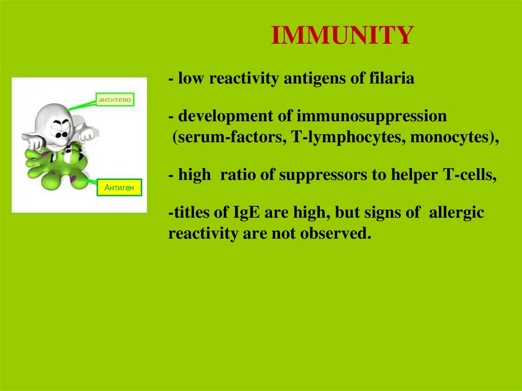
IMMUNITY- low reactivity antigens of filaria
- development of immunosuppression
(serum-factors, T-lymphocytes, monocytes),
Антиген
- high ratio of suppressors to helper T-cells,
-titles of IgE are high, but signs of allergic
reactivity are not observed.
20.
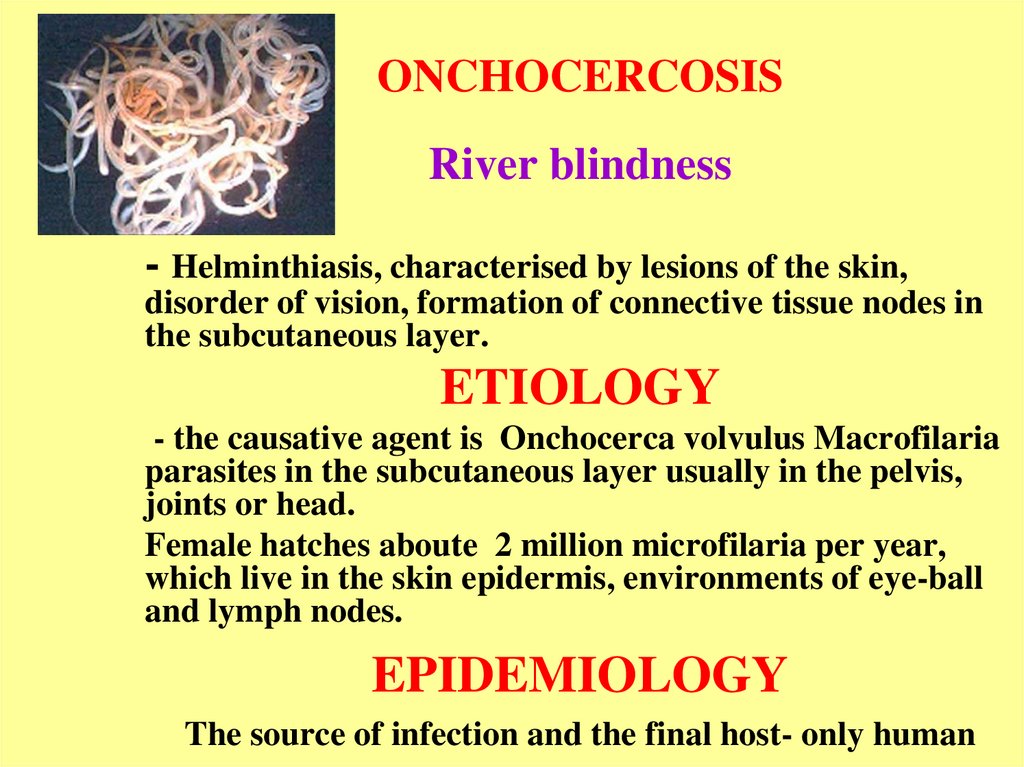
ONCHOCERCOSISRiver blindness
- Helminthiasis, characterised by lesions of the skin,
disorder of vision, formation of connective tissue nodes in
the subcutaneous layer.
ETIOLOGY
- the causative agent is Onchocerca volvulus Macrofilaria
parasites in the subcutaneous layer usually in the pelvis,
joints or head.
Female hatches aboute 2 million microfilaria per year,
which live in the skin epidermis, environments of eye-ball
and lymph nodes.
EPIDEMIOLOGY
The source of infection and the final host- only human
21.
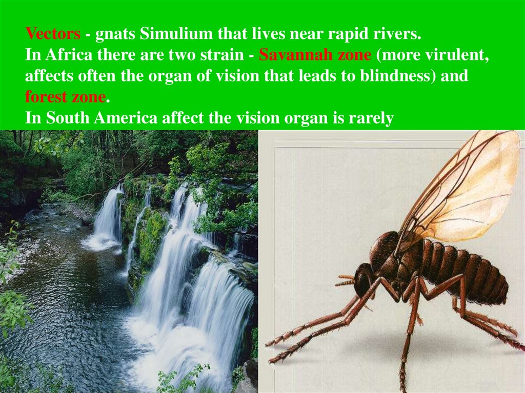
Vectors - gnats Simulium that lives near rapid rivers.In Africa there are two strain - Savannah zone (more virulent,
affects often the organ of vision that leads to blindness) and
forest zone.
In South America affect the vision organ is rarely
22.
PATHOGENESIS1. Mechanical effluence of adult parasites, around which
onchocercoma is formed (connective tissue node)
2. Toxic-allergic effects of mature parasites and it’s larvae
(especially dead worm)
3. Penetration of the larvae in the eyeball is manifested as
iritis or iridocyclitis («anterior uveitis») and/or chorioiditis
or chorioretinitis («posterior uveitis»), as keratitis,
conjunctivitis with subsequent development of gradual
sclerosis of the eyes, optic nerve atrophy and blindness
4. Parasitizing microfilaria causes dermatitis with lymph
swelling of the skin of genitals, lower extremities, and
elephantiasis
5. In the final stages depigmentation, atrophy, ulceration is
developed
23.
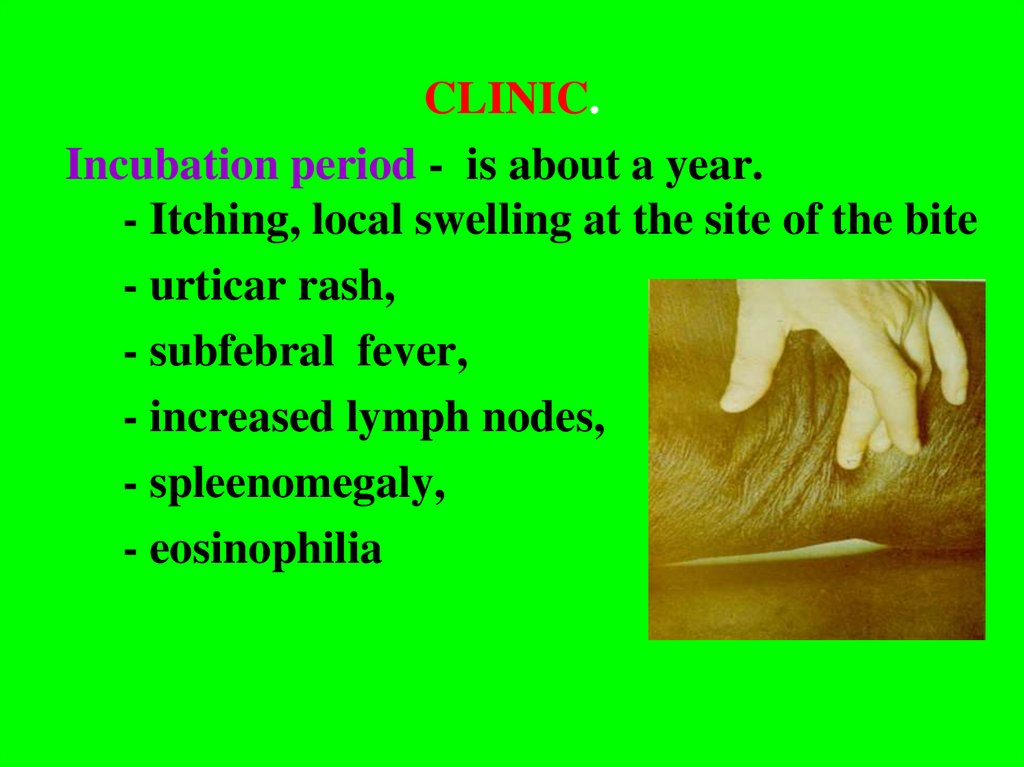
CLINIC.Incubation period - is about a year.
- Itching, local swelling at the site of the bite
- urticar rash,
- subfebral fever,
- increased lymph nodes,
- spleenomegaly,
- eosinophilia
24.
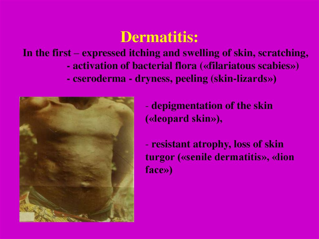
Dermatitis:In the first – expressed itching and swelling of skin, scratching,
- activation of bacterial flora («filariatous scabies»)
- cseroderma - dryness, peeling (skin-lizards»)
- depigmentation of the skin
(«leopard skin»),
- resistant atrophy, loss of skin
turgor («senile dermatitis», «lion
face»)
25.
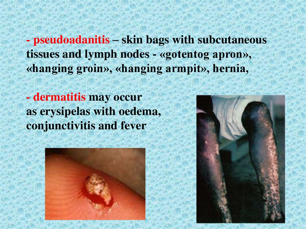
- pseudoadanitis – skin bags with subcutaneoustissues and lymph nodes - «gotentog apron»,
«hanging groin», «hanging armpit», hernia,
- dermatitis may occur
as erysipelas with oedema,
conjunctivitis and fever
26.
-Formation of onchocercoma - dense, mobile, painlessnodes with dead or live microfilaria.
-They have different sizes (from a pea to chicken
eggs), single or connected together in a thick capsule.
- In africans onchocercoma is localized below the belt
(scallops iliac bone, knee joints, side of the chest).
- In americans – upper part of the body (head, neck,
shoulders).
27.
-affection of lymphatic system lymphadenitis (groin and armpit),lymph oedema,orchitis, hydrocele,
elephantiasis of the lower limbs and
genitals
- microfilaria is detected in urine,
sputum, vaginal discharge, lymphatic
and blood circulation system, saliva,
cerebrospinal fluid, liver, kidneys, lungs,
spleen
Onchocercosis is a systemic disease
28.
Affection of eyes-corneal-conjunctivitis syndrome:
- pruritus, tearing, photophobia,
-blepharospasm.
-pointed keratitis, sclerosis, degeneration
and corneal ulcer.
-reduced visual function:
Iritis, iridotsyklitis, chorioretinitis.
Transparency of the conjunctiva
is lost,
the lens is cloudy, overgrown pupil.
-neuritis and optic nerve atrophy
and blindness.
29.
LOAOSIS(Calabar swelling disease)
Helminthiasis, characterised by the swelling of soft
tissues, affection of eyes and genital organs.
ETIOLOGY
Pathogen - Loa loa, adult worm parasites under the
conjunctiva of the eye and pericardium, microfilaria –
in the blood in afternoon.
30.
EPIDEMIOLOGYSource of invasion - man (sometimes monkeys)
Vectors - horse-flies of the
genus Chrysops that lives in
small water reservoirs.
Adult flies live in trees and
attack in the afternoon,
prefer people with dark skin.
31.
PATHOGENESIS – the same to other filariasisCLINIC
Incubation period - 4 months, more than a year.
1. Skin itching, rash, neuropathic pain, subfibrale fever,
hypereosinophilia.
2. Calabarien swelling on limited areas of the body (often
on the extremities), disappears slowly, painless, skin is
pale, hot, fossa is not remain.
3. Swelling, hyperemia of the conjunctiva, pain,
lacrimation. Helminth is visible by eyes.
4. Symptoms are correspond to the place of helminth
migration (dysuria, meningoencephalitis, neuritis,
nephrotic syndrome, hydrocele).
5. Abscesses around the dead worms.
6. Sometimes parasites visible under the skin and come
out through the skin.
32. LABORATORY DIAGNOSTICS 1. Detection of microfilaria in the blood smear and thick drops in the painted and unpainted
preparations with aquantitative assessment of microfilariemia.
M. Perstans
без чехлика
LOA LOA
Brugia malayi
с чехликом
O.volvulus без чехлика
33.
2. Detection of microfilaria in the skin sectionsreceived with sclera –corneal perforator
(onchocercosis).
34.
3. Detection of microfilaria in urine(W. and B.).
4. Ophtalmoscopic detection of microfilaria in the
front eye cavity (onchocercosis).
5. Detection of helminth under the conjunctiva directly
(loaosis).
6. Маzоtti-test with ditrasune (except for loaosis).
7. Immunological methods (CBR, RIHA).
35.
TREATMENTDietylcarbamasepine - is effective in acute and chronic
stage, in latent filariasis
6 mg /kg /day (after meal) - 12 days (from 3 to 6 mg /
kg / day).
In loaosis - on the first day - 1\2 of doses, gradually
increasing to 0.1 (3-4 times) - 2-3 weeks.
Onchocercosis:
Dietylcarbamasepine (initial dose reduced) -12 days
Antripol (suramin)- 10% - 5ml - 1st day
10% - 10 ml - 2nd day
10% - 10 ml -1 times in 7 days 5-7 weeks
Ivermectine (mektisane) 150 mg\kg 1 time in 6 months.
36.
PREVENTION1. Straggle with the intermediate hosts
2. Improvement of the source of the invasion: therapy of sick
people
3. Sanitary-hygienic measures on improvement of settlements
(water, sewerage, shower and other).
4. Individual prevention - protective clothing at risk groups.
5. Health education of the population (not pollute the water
with feces, not to swim and others).
6. Sanitary supervision over natural reservoirs
37.
Thank you for attention!Stay healthy!