


 Медицина
Похожие презентации:


Ultrasound is
1.
2. Plain films
Plain films still remain the mainstay ofradiological investigation of the skeletal
system. Views should always be obtained in
two projections.
3.
4. Ultrasound
Ultrasound is utilized for the evaluation of:neonatal hip for congenital dislocation
soft-tissue lesions, abscesses and masses
joint effusion
5.
6. CT scan
CT aids:assessment of bone tumors prior to surgery
evaluation of certain fractures, such as the
acetabulum and subtalar joint
study of the spinal column
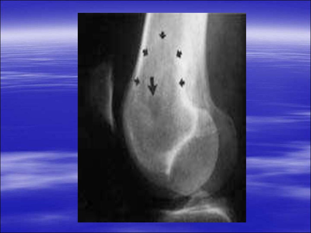
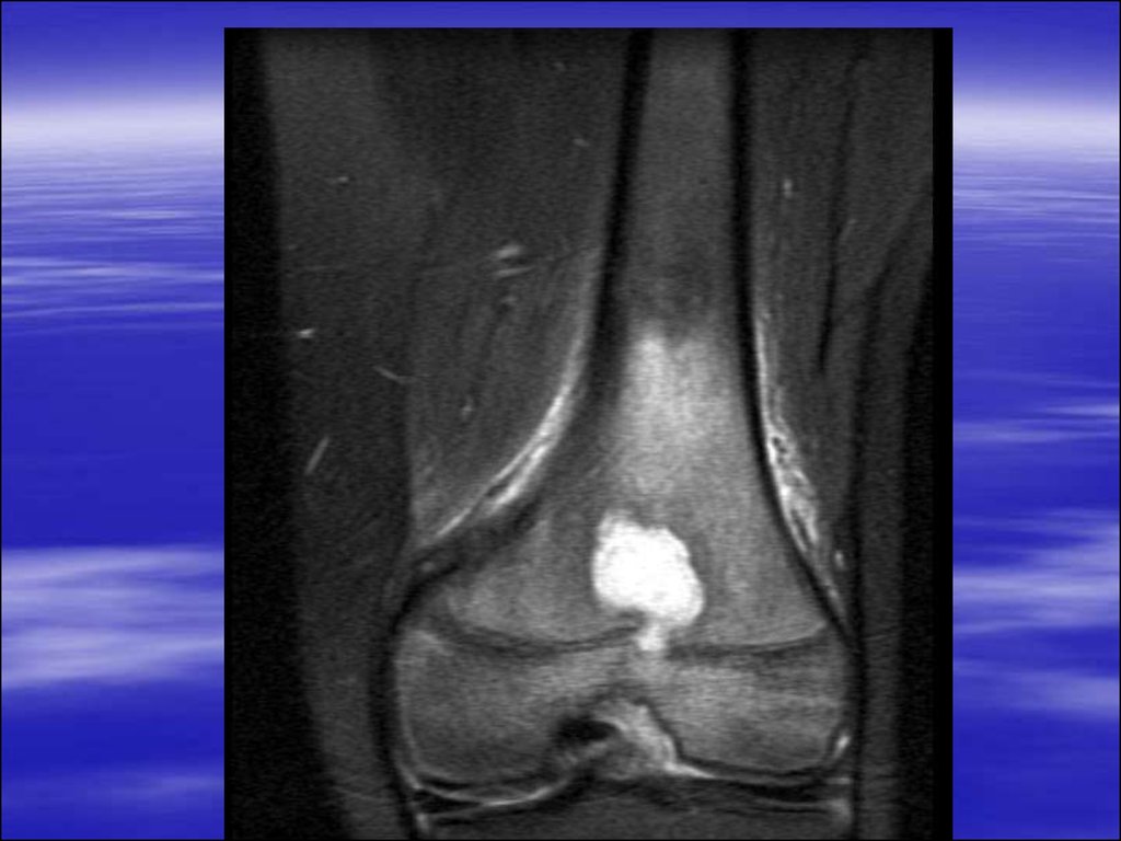
7. Aneurysmal bone cyst
8.
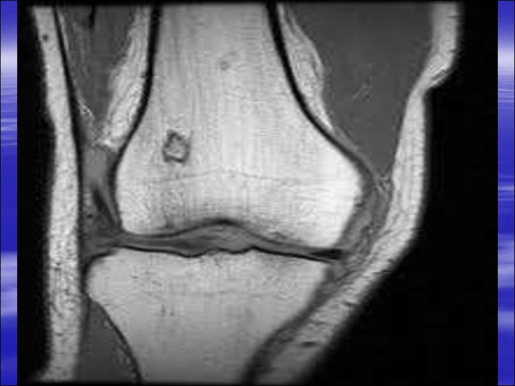
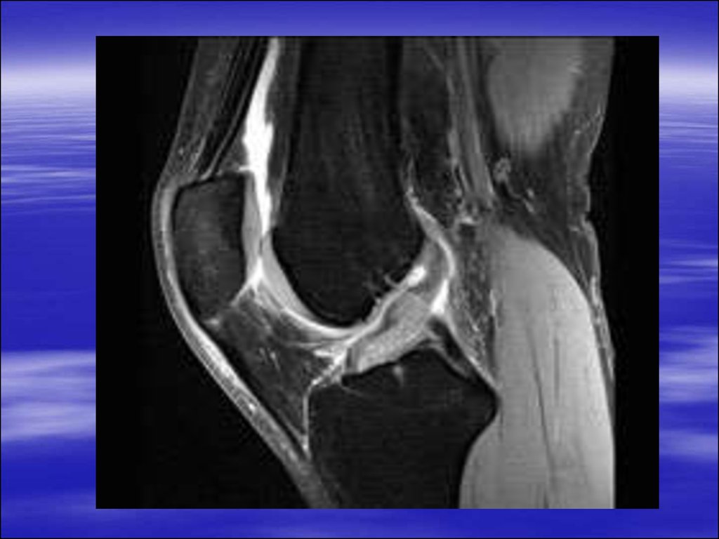
9. MRI
MRI assists the:investigation of bone tumor
soft tissue masses
the spinal column and joints
10.
11.
12. Isotopes scan
Technetium 99 phosphonate compoundsaccumulate in bone several hours after
intravenous injection of the isotope; principally
used for:
detection of osteomyelitis and other
musculoskeletal soft-tissue inflammatory changes
metastatic bone lesions: changes are seen much
earlier than plain films
staging tumors such as breast carcinoma or
bronchial carcinoma
functional bone abnormality: Paget’s disease
13. An isotope bone scan showing hot spots in the left foot and in the ribs, suggestive of metastases.
14. Arthrography
In this procedure, contrast and air areinjected into joints such as the knee, hip,
elbow, shoulder, wrist and
temporomandibular joints to diagnose
loose bodies
ligamentous abnormalities
cartilaginous abnormalities
15.
16. the left shoulder in external rotation
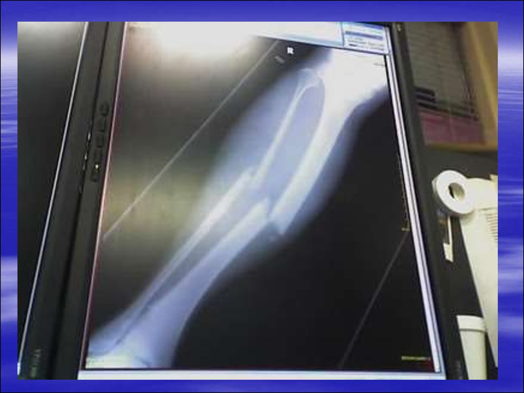
17. Skeletal trauma
FractureFracture is defined as complete or incomplete
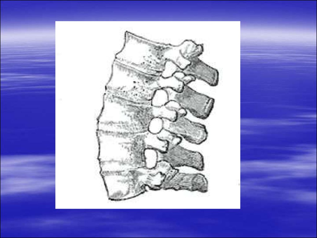
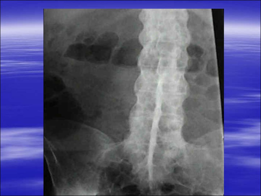
disruption in the continuity of bone.
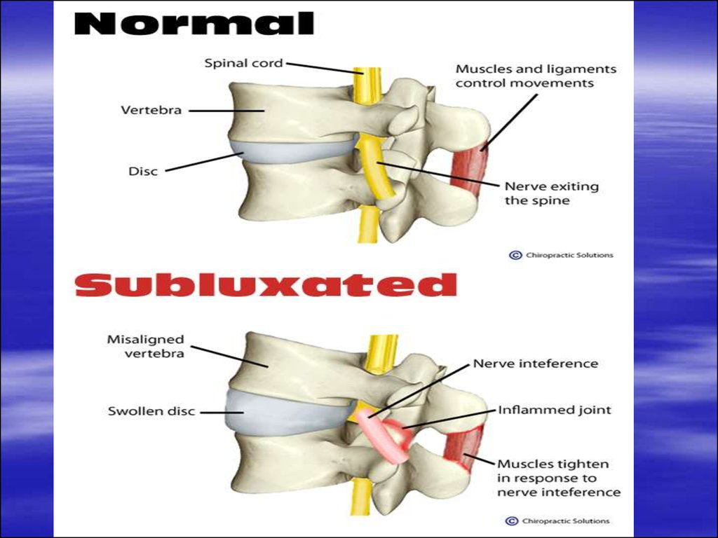
Dislocation
Dislocation is defined as the complete disruption
of the alignment of the articular surfaces of the
joint.
Subluxation
Subluxation is defined as the incomplete
disruption of the a ligament of articular surfaces.
18.
19.
20.
21. Evaluation of fracture
Complete radiographic evaluation of fractureshould include:
site and extent of the fracture
type of fracture
alignment of the fractured fragment
direction of fracture line
dislocation or Subluxation of the adjacent joint
associated abnormalities
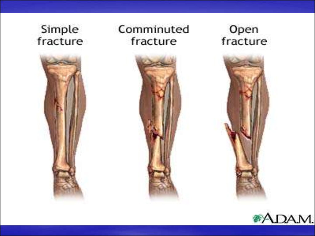
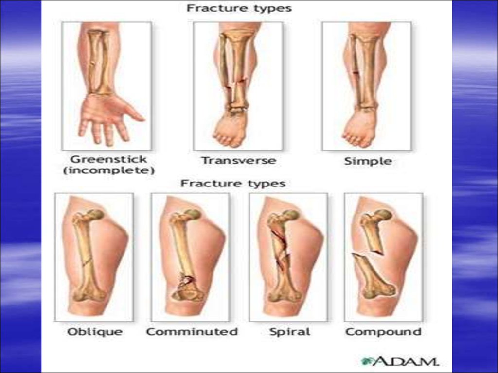
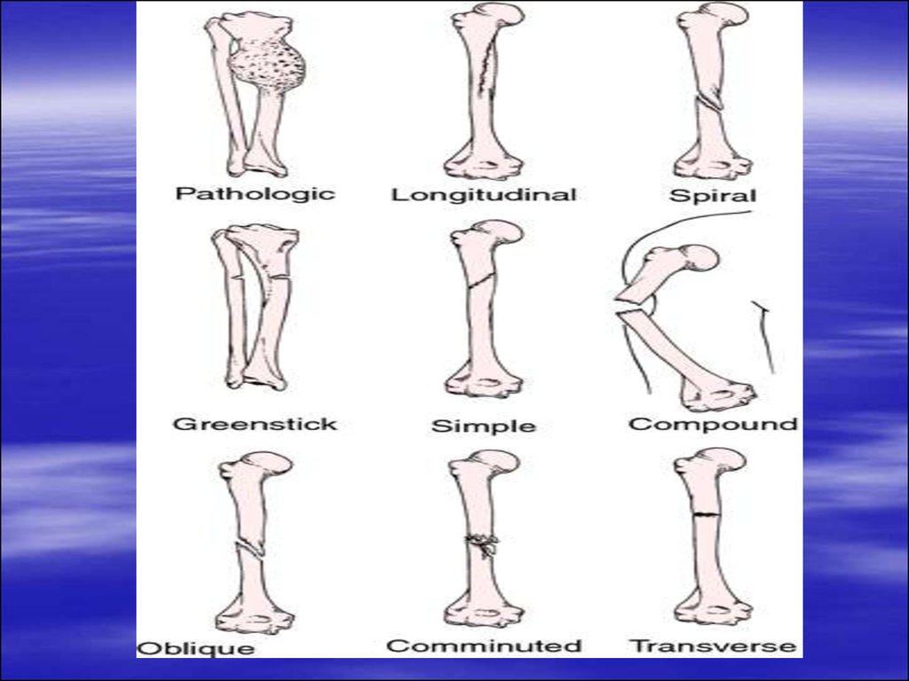
22. Types of fractures
based on the fracture line and the number offractured fragments fractures are classified into:
–
–
simple fracture: here single fracture line is seen with
two fracture fragments
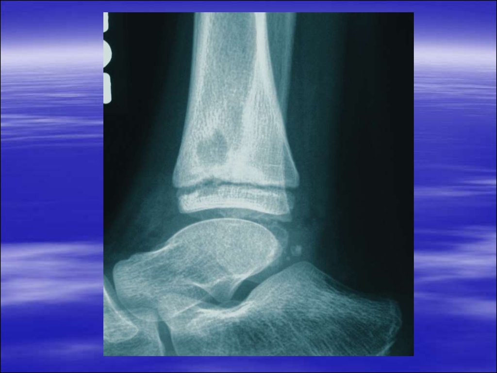
comminuted fractures: here multiple fracture
fragments are seen
based on whether the fracture is exposed to the
external surface or not, the fractures can be
classified into:
–
–
closed fractures: here there is no communication of
the fracture with the exterior
open fractures: here the fractured fragments are
exposed to the exterior trough a skin wound
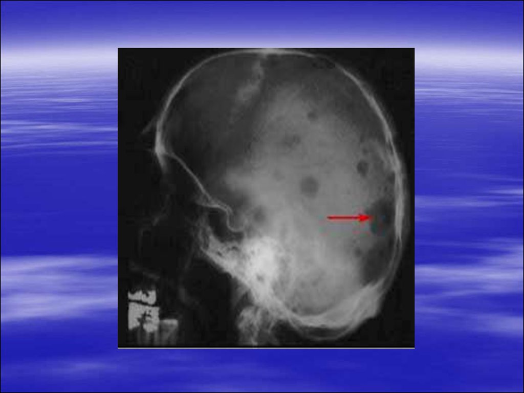
23.
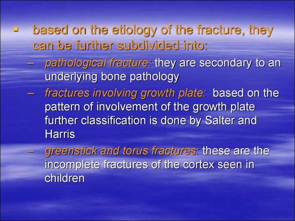
based on the etiology of the fracture, theycan be further subdivided into:
– pathological fracture: they are secondary to an
underlying bone pathology
– fractures involving growth plate: based on the
pattern of involvement of the growth plate
further classification is done by Salter and
Harris
– greenstick and torus fractures: these are the
incomplete fractures of the cortex seen in
children
24. Types of fracture lines
horizontaloblique
spiral
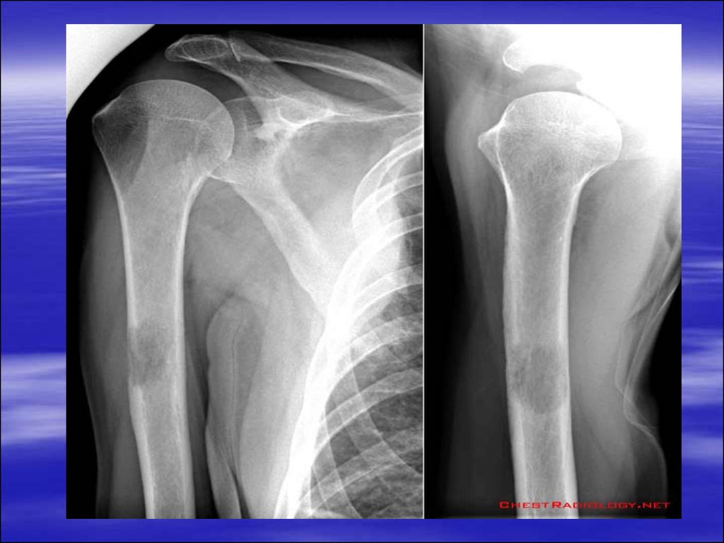
vertical
25.
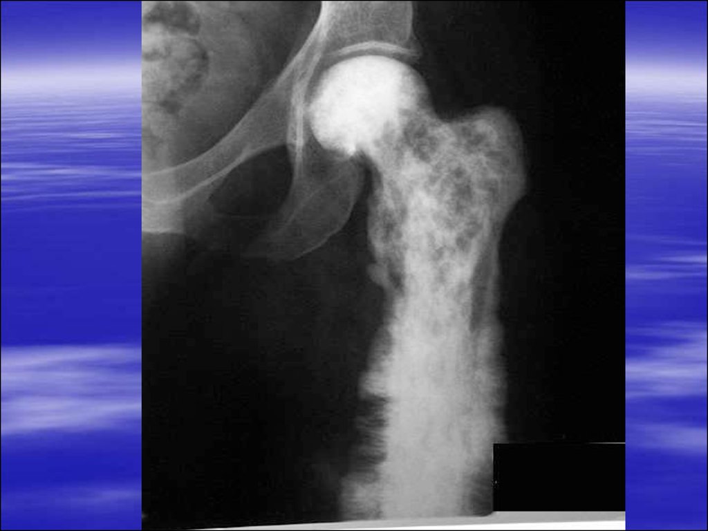
26.
27.
28.
29.
30.
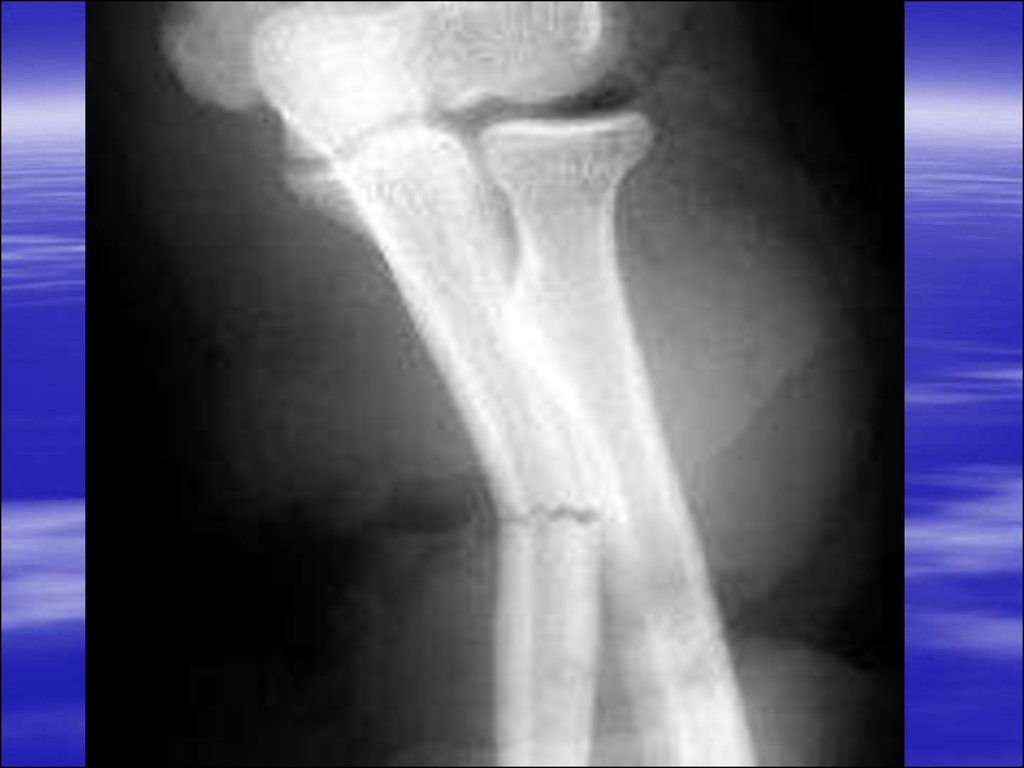
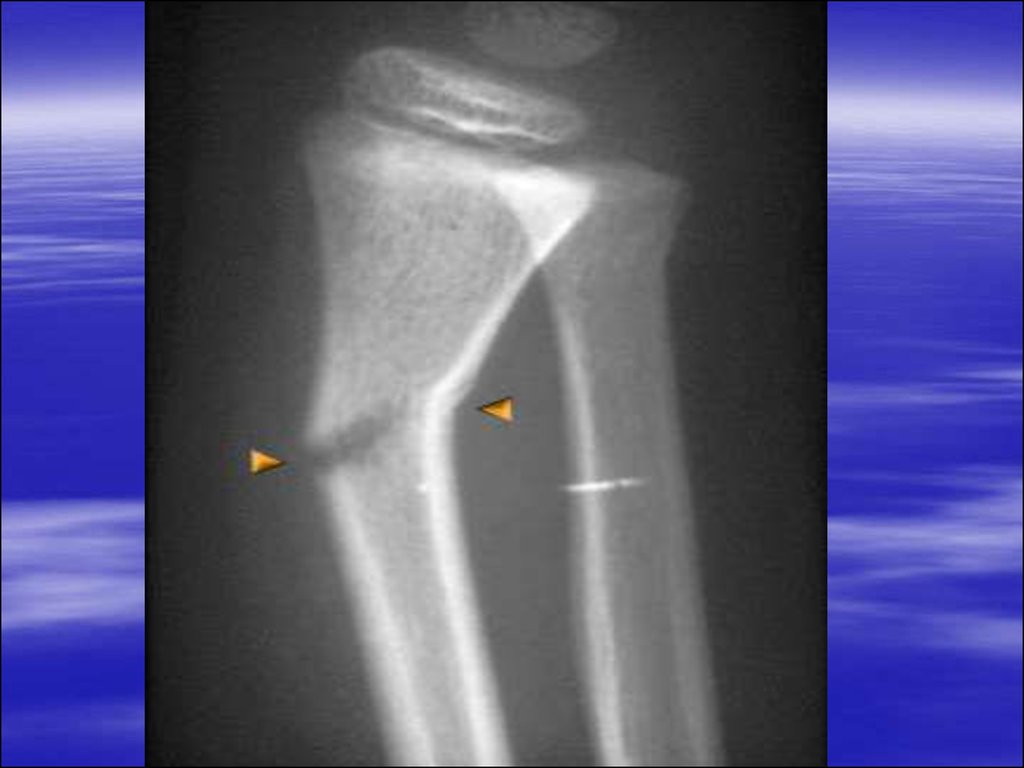
31. Oblique fractures of the radius and ulna.
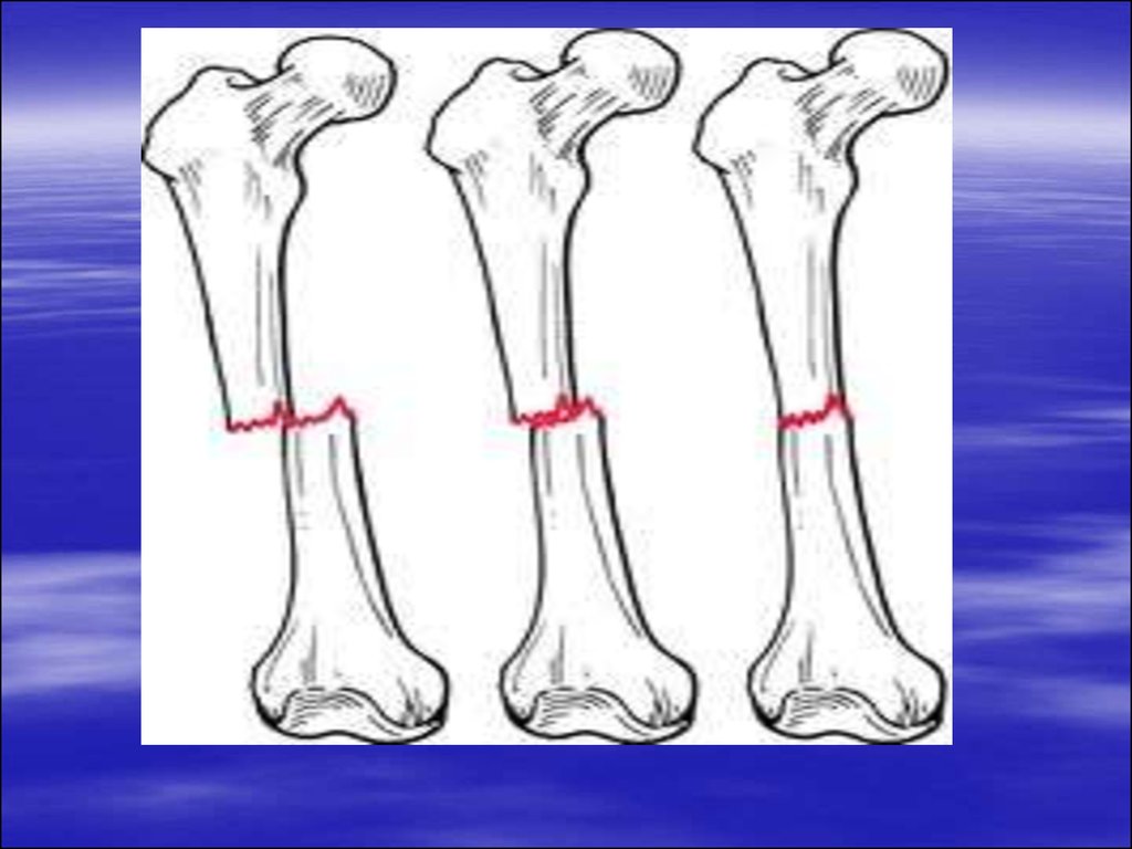
32. Types of displacement of fractured fragments
medial displacementlateral displacement
medial angulation (or lateral angulation of distal
fragment-valgus configuration)
lateral angulation (or medial angulation of distal
fragment-varus configuration)
internal rotation
external rotation
overriding with foreshortening (bayonet apposition)
distraction
33.
34.
35.
36.
37.
38.
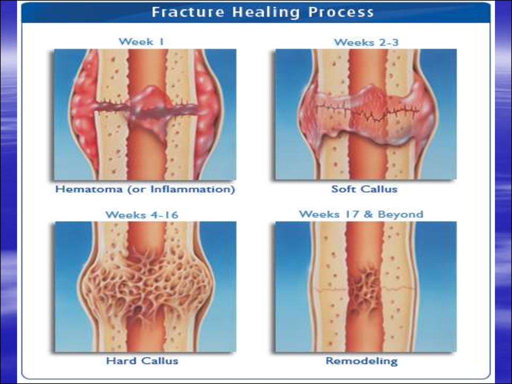
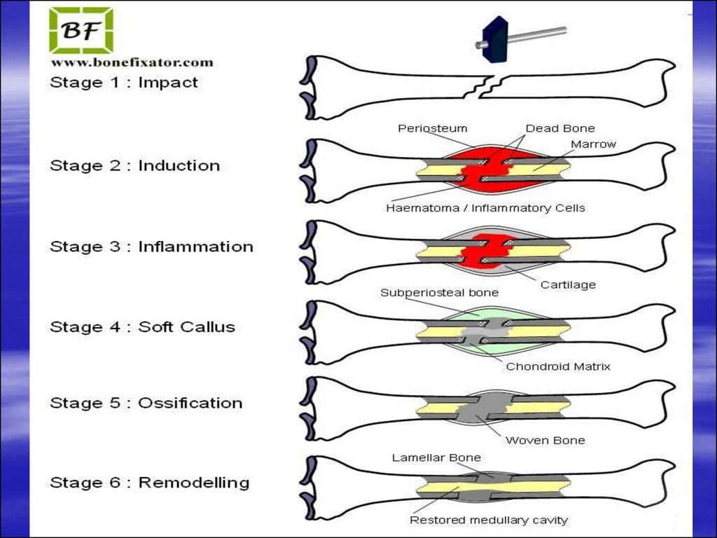
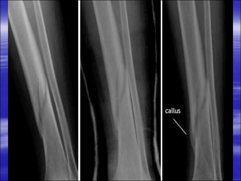
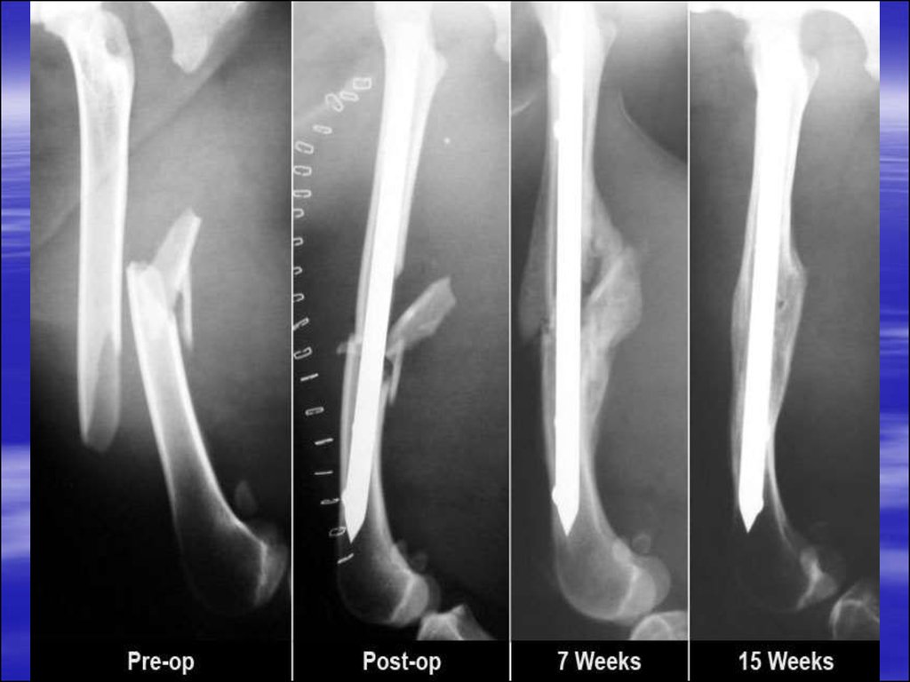
39. Mechanism of fracture healing
primary union – this type of healing is seenin undisplaced and perfectly reduced
fractures and the healing occurs by
endosteal callus formation
secondary union- this type of healing in
displaced fractures and the healing is by
periosteal callus formation
40.
41.
42.
43.
44. Complications of bone healing
mal-union – this is the most commoncomplication of fracture healing; here the fracture
healing occurs in the mal-aligned fracture
fragments
delayed union- the fracture healing is delayed for
16-18 wks due to underlying infection or
improper immobilization
non union – no healing will be noted in the
fractured fragments and the margins are
sclerosed
disuse osteoporosis and reflex sympathetic
dystrophy syndrome
45.
myositis ossificans: due to prolongedimmobilization and soft tissue ossification
mainly around the hip region
osteonecrosis: interruption of the vascular
supply leads to avascular necrosis; this
complication is common with fracture of
scaphoid and fracture neck of femur
injury to major blood vessels
growth disturbance
post traumatic arthritis
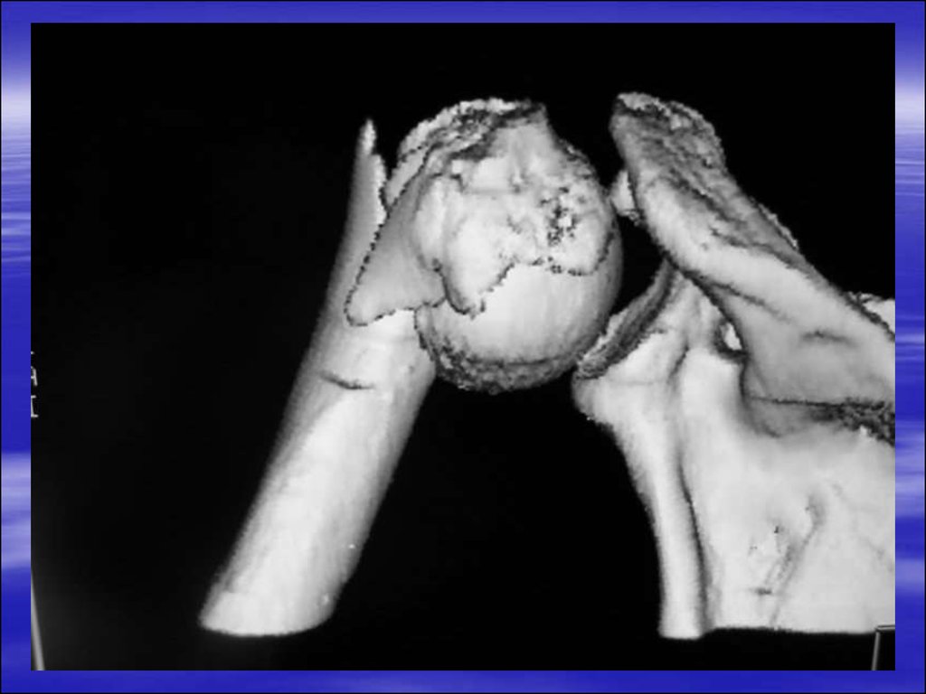
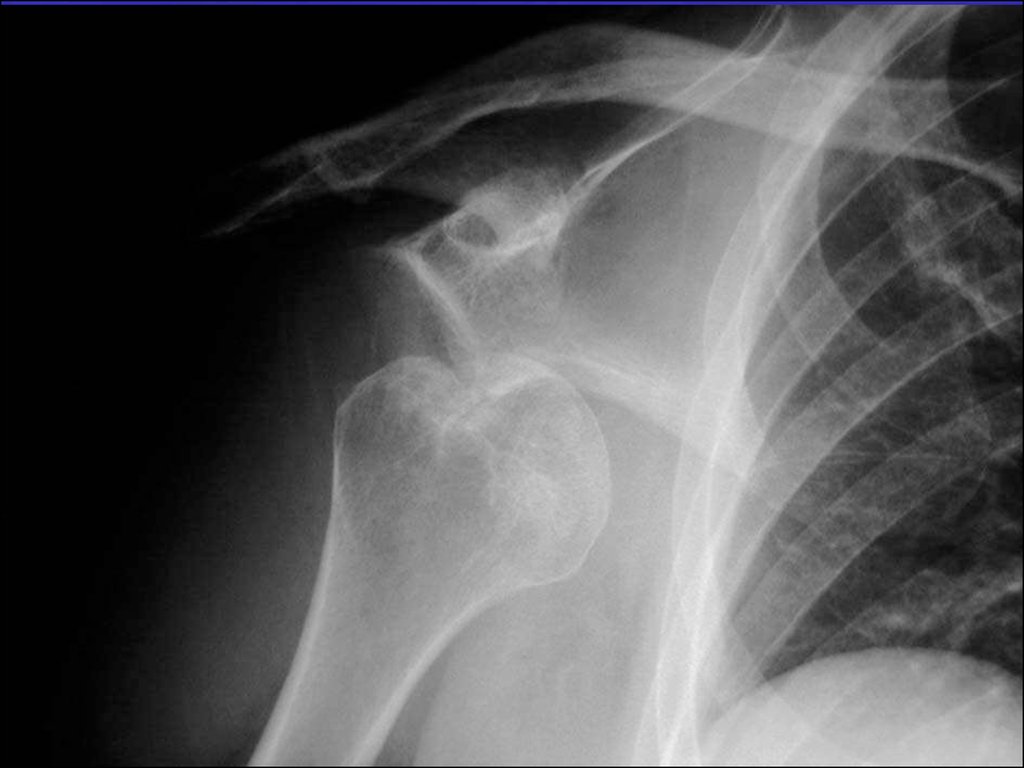
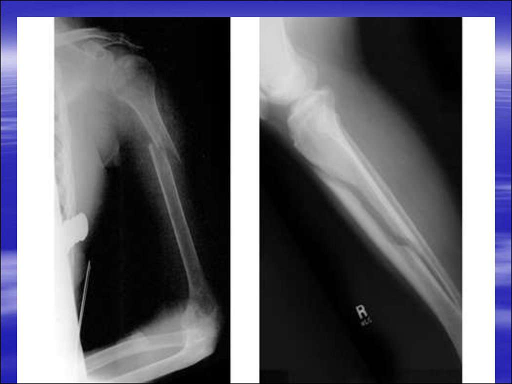
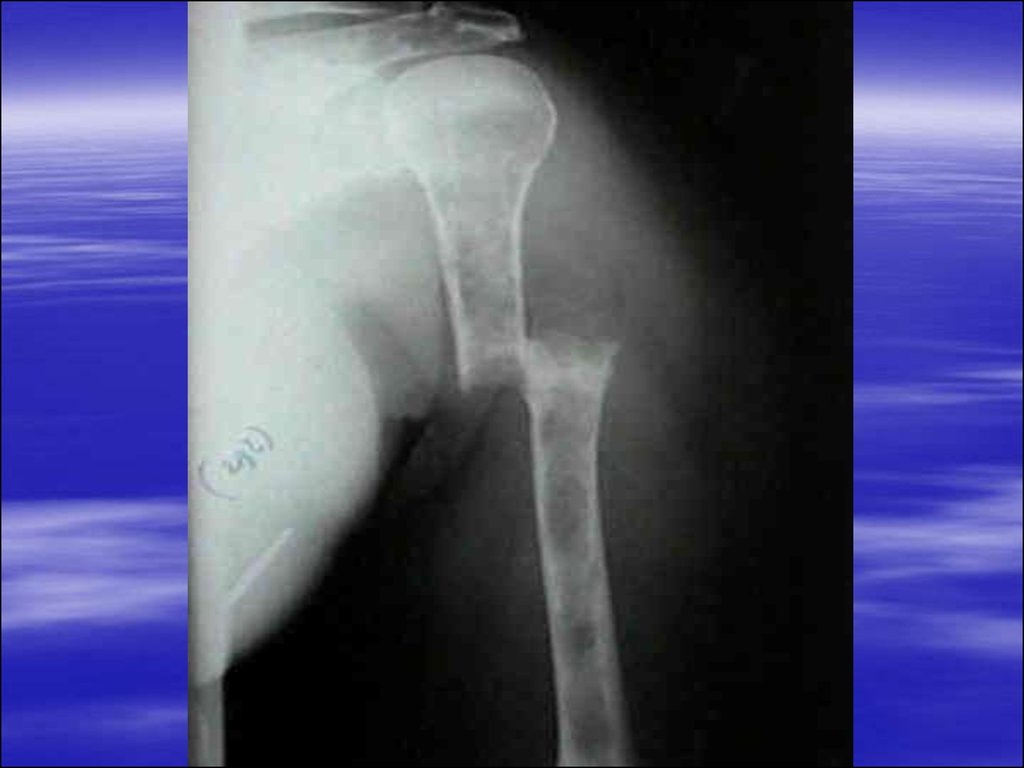
46. Glenohumeral dislocations
Anterior dislocationThis is the most common type of glenohumeral dislocation.
Humeral head is dislocated anterior to
glenoid fossa.
Force which predisposes to anterior
dislocation is the combination of abduction,
extension and external rotation.
The bone lesions associated with recurrent
anterior dislocations of the shoulder
47.
Posterior dislocationless common
force predisposing to the posterior
dislocation is – adduction, flexion and
internal rotation
humeral head is displaced posterior to
glenoid fossa
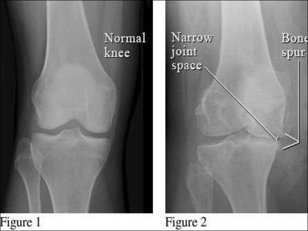
48. Osteoporosis
Osteoporosis is a condition in which there isa reduction of bone mass.
Presentation
asymptomatic
bone pain
skeletal fractures
vertebral compression fractures
49. Radiological investigations
plain filmsCT scan
Radioisotope scan
50. Radiological features
decrease in the number of trabeculaecoarse striations
the vertebral bodies appear lucent with thin
cortical lines
biconcave appearance (“cod fish” vertebrae)
vertebral wedging and collapse
kyphosis
fractures of the peripheral skeleton,
including femoral neck fractures, commonly
occur even after minor trauma
51. Causes of generalized osteoporosis
senile osteoporosispostmenopausal
steroid therapy
immobility (prolonged bed rest)
endocrine (Cushing’s disease)
multiple myeloma
nutritional deficiency syndrome( scurvy,
malnutrition, chronic liver disease,
malabsorption syndrome)
52.
53.
54. Ankylosing spondylitis
Ankylosing spondylitis, a progressiveinflammatory disease, usually affects young
adult males, often with a family history of the
disease.
Presentation
repeated attacks of backache and stiffness
anorexia and weight loss
55. Radiological features
On plain films the following features may beseen:
sacroiliac joints: the earliest changes begin
in the sacroiliac joints with symmetrical
blurring and poor definition of joint margins;
later, erosion and bony sclerosis lead to
tendency for complete sacroiliac joint fusion;
both joints are commonly affected
56.
spinal changes: the entire spine may be involvedbut changes usually commence in the lumbar
region and progress upwards to involve the
thoracic and cervical spine; the features most
commonly noted are: squaring of the vertebral
bodies due to new bone formation in the anterior
vertebral bodies, and filling in of the normal
anterior concavity by longitudinal ligamentous
calcification; calcification of the lateral and anterior
spinal ligaments to produce the classical “bamboo
spine”
peripheral joint involvement: an erosive
arthropathy may accompany ankylosing
spondylitis, the hips being the commonest joints
involved
57. Complications
upper-lobe fibrosisaortic incompetence: from an aortitis of the
ascending aorta
inflammatory bowel disease: a colitis resembling
Crohn’s disease or ulcerative colitis
atlanto-axial subluxation
fractures: spinal rigidity causes increased
susceptibility to trauma
ventilatory failure: due to restrictive chest
movements and ankylosis of the costovertebral
joints
iritis
58.
59.
60. Osteomyelitis
infection of the boneroutes of spread may be haematogenous or direct
spread from the infected joint or infected wound
staphylococcus is the most common organism
in infants the site of predilection is metaphyseal
with epiphyseal extension; in children it is
metaphyseal while in adults it is epiphyseal
There are two types of osteomyelitis: acute and
chronic.
61. Acute osteomyelitis
presents with an acute episode of pain andreduced functioning of the part with the
systemic ill-health
more common in boys
62. Imaging features
initial radiographs are normal as bone changesare not visible upto 10-14 days of infection; Tc99
radionuclide scan shows increased uptake after 23 days
MRI also picks up early osteomyelitis where in the
normal marrow signal intensity is lost in T1
weighted images due to oedema with soft tissue
swelling
Typically acute osteomyelitis affects metaphysis
of long bones, usually femur and tibia
– features are soft tissue swelling with blurring of fat
planes
– focal osteopenia (rarefaction) of the bones seen in the
metaphyses with periosteal reaction
63.
64.
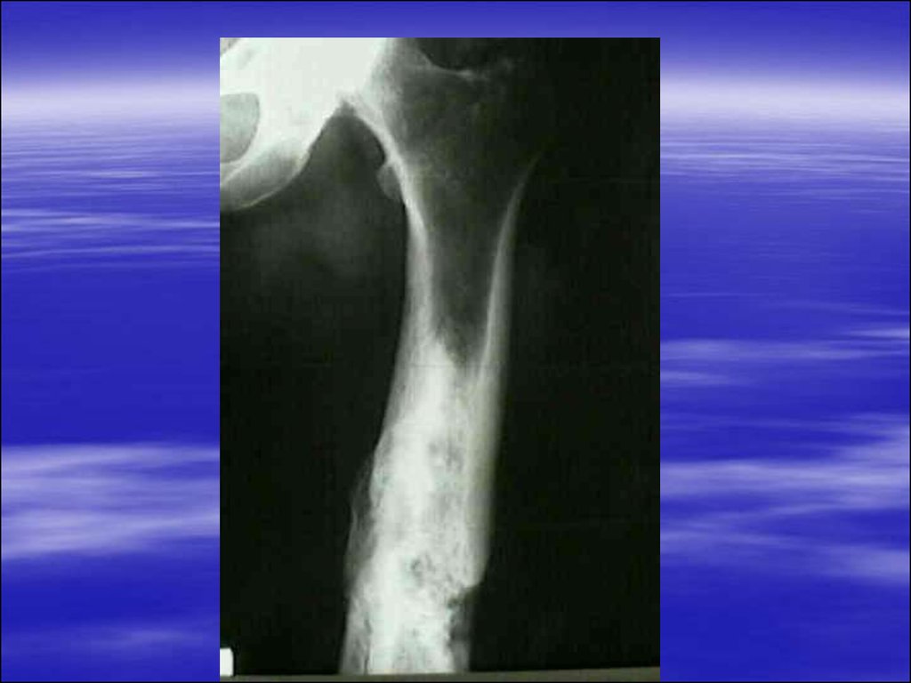
65. Chronic osteomyelitis
sequelae of acute osteomyelitisin chronic osteomyelitis, bone becomes
thickened and sclerotic with loss of
differentiation between cortex and medulla
66. Imaging features
2-6 weeks after acute infection, there isprogressive destruction of cortical and medullary
bone with increased endosteal sclerosis, indicating
reactive new bone formation and periosteal
reaction
In 6-8 weeks, “sequestra” which are areas of
necrotic bone become apparent; they appear more
sclerotic (more dense) because of the relative
decrease in density in the adjacent bone and lack
or remodeling
67.
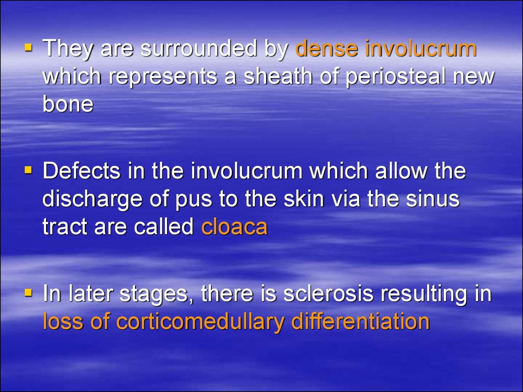
They are surrounded by dense involucrumwhich represents a sheath of periosteal new
bone
Defects in the involucrum which allow the
discharge of pus to the skin via the sinus
tract are called cloaca
In later stages, there is sclerosis resulting in
loss of corticomedullary differentiation
68.
69.
70. CT scan:
it demonstrates changes in subacute orchronic osteomyelitis well, especially those
related to cortical bone or periosteum
sequestra, as on conventional films, are
shown as areas of dense or right attenuation
spicules of bone lying in areas of osteolysis
cloacae, periostitis and local soft tissue
masses are shown
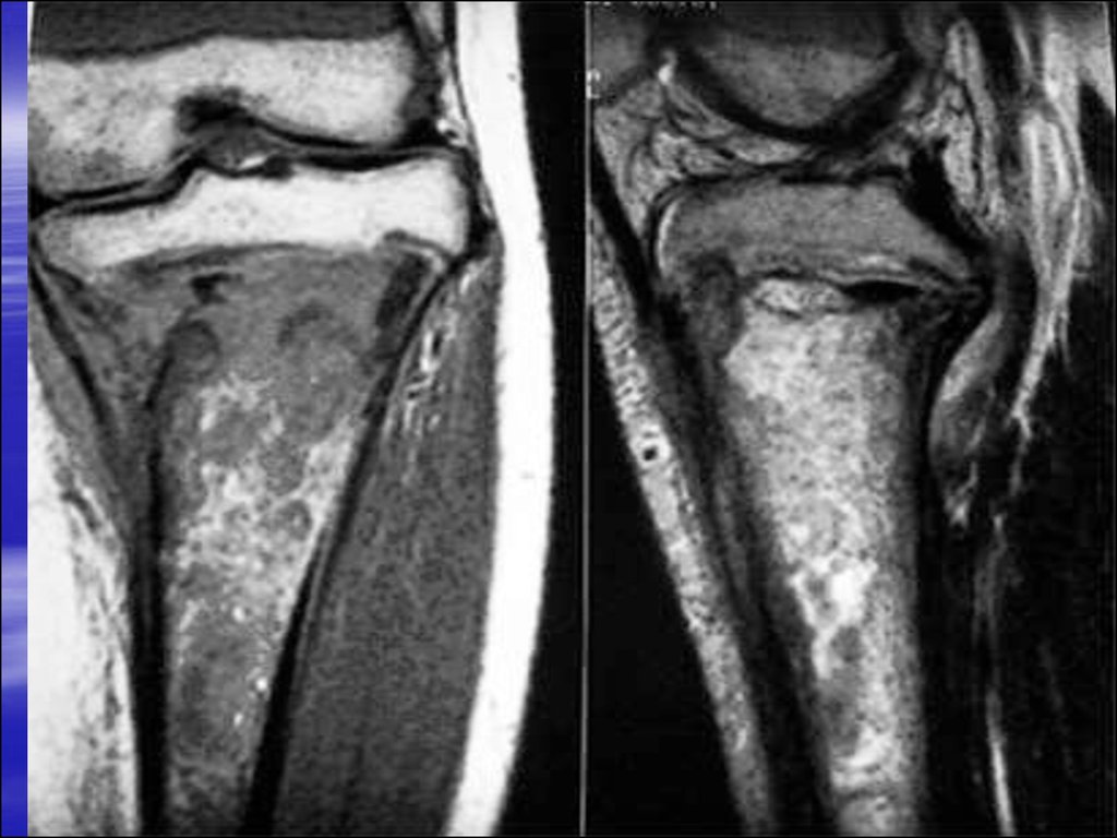
71. MRI
demonstrates osteomyelitis as early asisotopic scanning and when available is the
modality of choice in the diagnosis of
musculoskeletal infection
demonstrates soft tissue edema
ischemia
destruction of cortex or marrow can be seen
at early stage
soft tissue extension of pus through cloacae
and para-osseous abscesses may be seen
72.
73. Special forms of osteomyelitis
Sclerosing osteomyelitis of Garremanifested by the gross sclerosis in the
absence of apparent bone destruction
bone appears thickened due to periosteal
new bone formation and loss of
corticomedullary differentiation
74.
75. Brodie’s abscess
sub acute infectionusually seen in the cancellous tissue near
the end of long bone
well-circumscribed areas of bone
destruction, which is surrounded by intense
sclerosis
76.
77.
78. Multiple myeloma
Multiple myeloma is primary malignanttumor of bone marrow, in which there is
infiltration of the marrow-producing areas of
skeleton by a malignant proliferation of
plasma cells.
79.
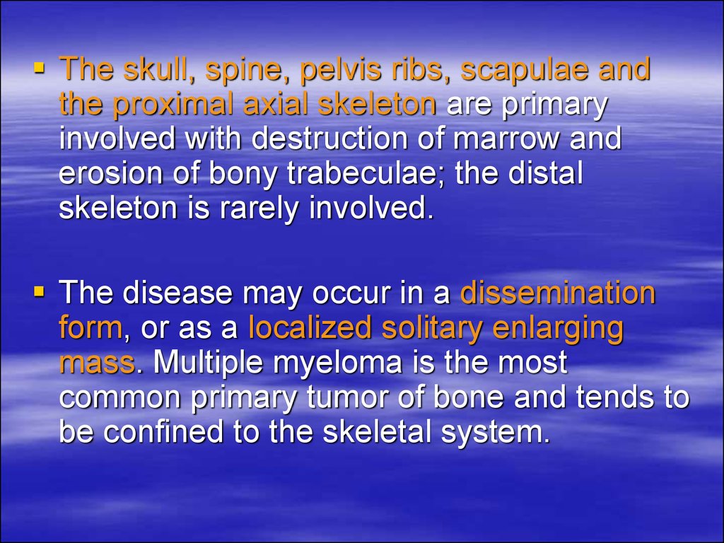
The skull, spine, pelvis ribs, scapulae andthe proximal axial skeleton are primary
involved with destruction of marrow and
erosion of bony trabeculae; the distal
skeleton is rarely involved.
The disease may occur in a dissemination
form, or as a localized solitary enlarging
mass. Multiple myeloma is the most
common primary tumor of bone and tends to
be confined to the skeletal system.
80.
Presentationa male predominance, usually in the over-40
age group
bone pain
backache
vertebral body collapse
pathological fracture
Bence-Jones proteinuria
81. Radiological features
generalized osteoporosis with a prominence of thebony trabecular pattern, especially in the spine,
resulting from marrow involvement with myeloma
pathological fractures are common
compression fractures of the vertebral bodies,
indistinguishable from those of senile osteoporosis
scattered “punched-out” lytic lesions with welldefined margins, those lying near the cortex
produce internal scalloping
bone expansion with extension through the cortex,
producing soft-tissue masses
82. Complications
– pathological fractures that heal with abundantcallus
– hypercalcaemia secondary to excessive bone
destruction
– renal failure may result from a combination of
amyloid deposition, hypercalcaemia and tubular
precipitation of abnormal proteins
– increased incidence of infections such as
pneumonia
– hyperuricaemia and secondary gout
83.
84.
85.
86. Bone metastases
Bone metastases are the most commonmalignant bone tumors. Metastases
disseminate mainly to marrow-containing
bones, therefore they are more commonly
found in the axial skeleton. Generally,
spread distal to the knee and elbow is less
likely than the proximal skeleton.
87.
Any primary tumor may metastases to bone,but the most frequent to do so are:
breast: high incidence of bone deposits,
usually lytic in nature but may be sclerotic or
mixed; the commonest cause of sclerotic
deposits in females
prostate: almost always sclerotic, lytic
deposits being rare; the commonest cause
of sclerotic deposits in a male
88.
lung: lytic deposits; peripheral deposits inthe hands and feet are rare, but if present
are likely to be form a bronchial carcinoma
kidney, thyroid: lytic and can be highly
vascular with bone expansion
adrenal gland: predominantly lytic
89.
Presentationbone pain
pathological fracture
soft-tissue swelling
staging or during follow-up of primary
tumors
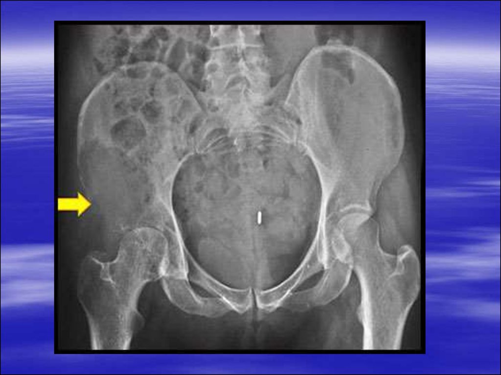
90. Radiological features
Lytic depositsDestruction of bone detail with poor
definition of margins and associated
pathological fractures are the principal
features. Periosteal reactions are rare
compared to primary malignant tumors.
91.
Sclerotic depositsShow as an area of ill-defined increased
density with subsequent loss of bone
architecture. Vertebral secondaries may
feature sclerotic pedicles. With multiple
lesions, a diagnosis of metastases is almost
certain. Isotope bone scanning is more
sensitive than plain films (localized areas of
increased uptake: hot spots).
92. Differential diagnosis
Paget’s disease (sclerotic areas)Multiple myeloma (lytic areas)
Primary tumor
Infection or osteomyelitis
93.
94.
95.
96.
97.
98.
99. osteolytic-sclerotic bone
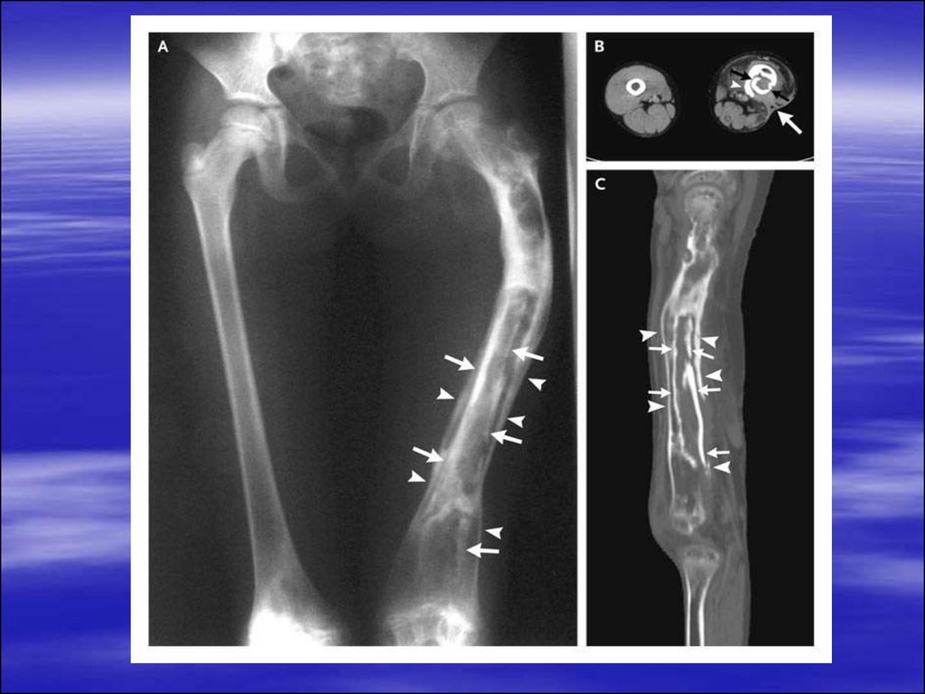
100. Paget’s disease
Paget’s disease is a common disorder ofbone architecture, of known aetiology, which
occurs with increasing frequency after
middle age. It is characterized initially by
bone deposition results in bone expansion
and abnormal modeling.
101.
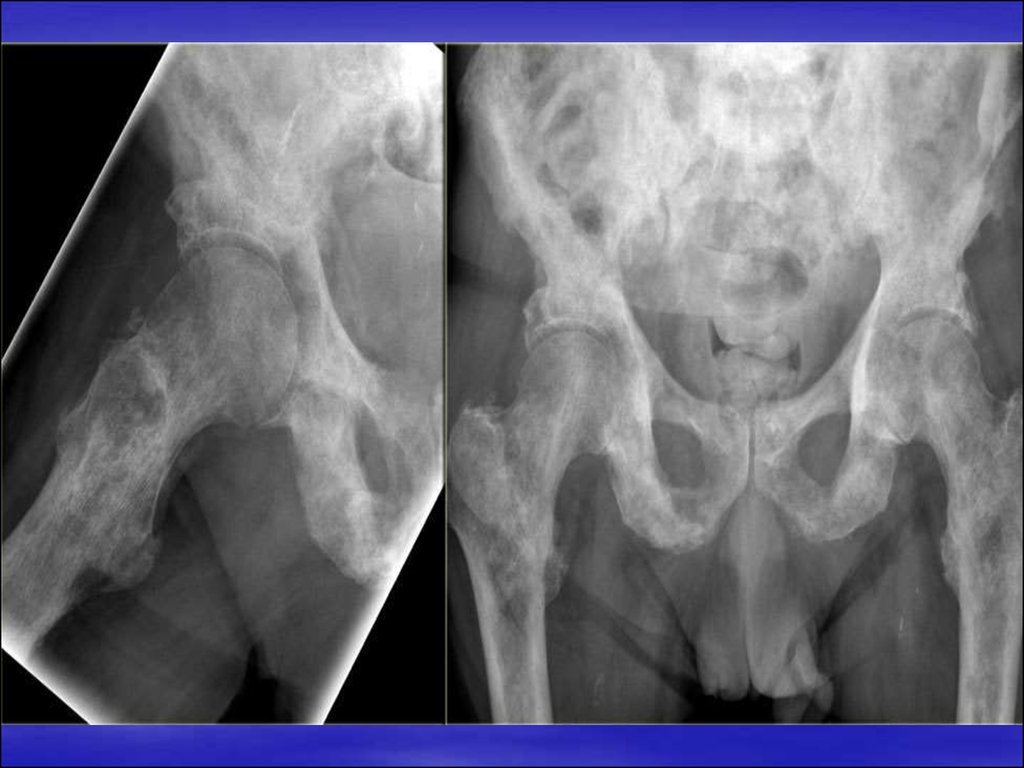
PresentationAny bone may be affected.
Skull: initially a large area of well defined
bone loss may be seen; later, generalized
sclerosis with diploic thickening produces a
characteristic “cotton wool” appearance;
they may be an increase in the size of the
head
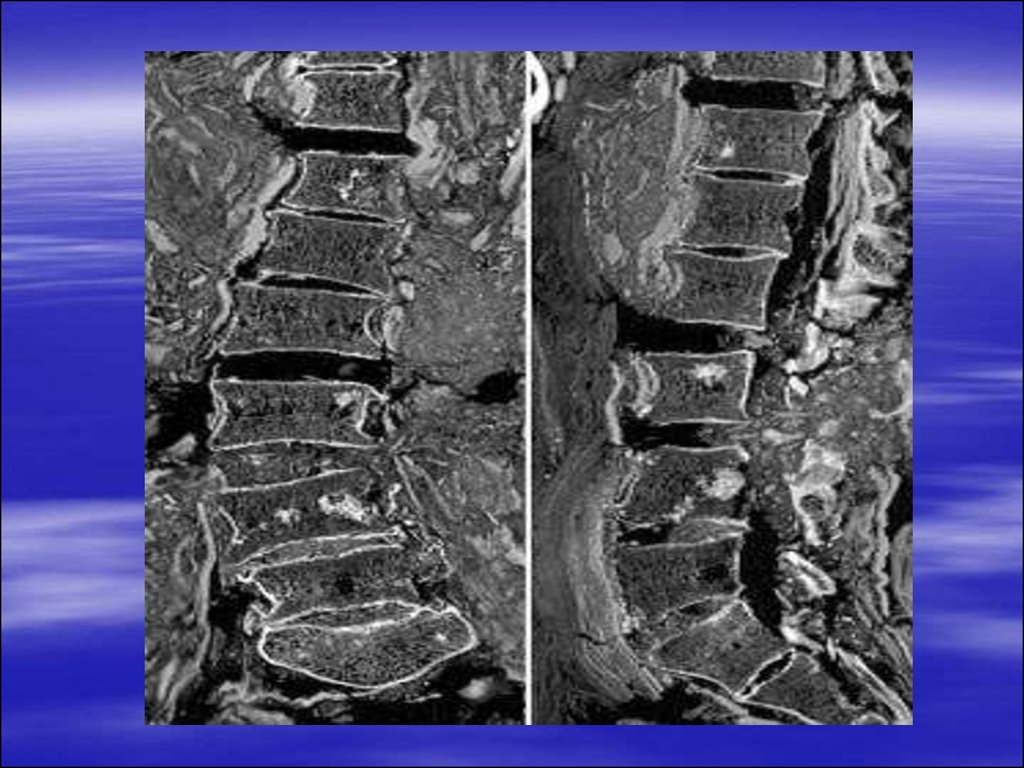
102.
Spine: most commonly involves a singlevertebra with sclerosis, altered trabecular
pattern and enlargement of the vertebral
body
Pelvis: frequently affected with coarsened
trabecular pattern, cortical thickening and
enlargement of the pubis and ischium
Long bones: widening of bone with
deformities, bowing of the tibia and
incomplete fractures because of bone
softening
103. Complications
pathological fractures: tend to be sharplytransverse
pseudofractures: incomplete fractures found on
the convex surfaces of bowed bones
malignant degeneration: in widespread Paget’s
disease there is an increased incidence of
malignant bone tumors, especially osteogenic
sarcoma
cardiovascular: increased shunting of blood in
involved bone may cause high output failure,
although this is rare
neurological: nerve entrapment by bone expansion
104.
105.
106.
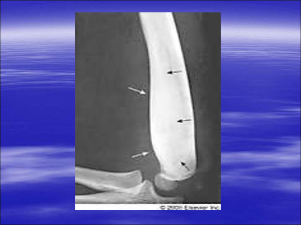
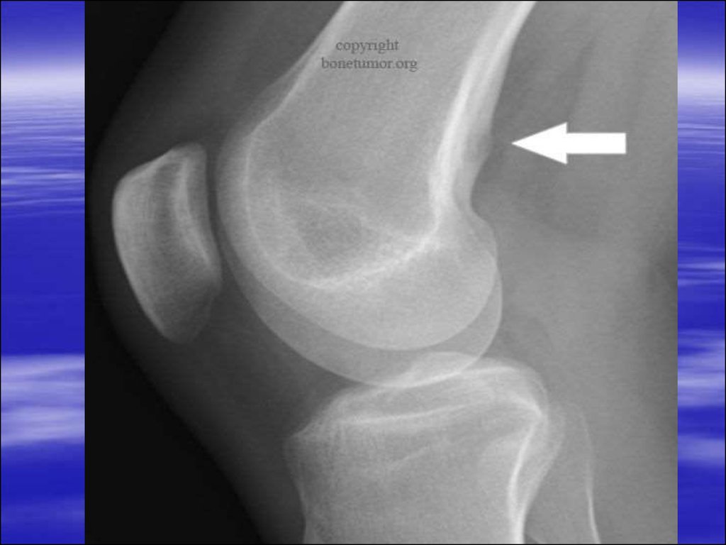
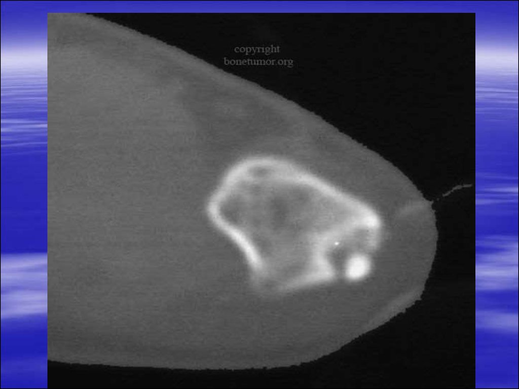
107. Osteoid osteoma
Age: 2nd and 3rd decaySite: More common in the long bones in
metaphyses or diaphysis of tubular bones
like femur and tibia. Classical clinical
presentation of sever bone pain aggravated
in the night and relieved by aspirin.
108. Radiological appearances:
round or oval lesion with a sclerotic marginthe radiolucency consists of a small dense
opacity known, as the nidus
the size of lesion is upto 2.5 cm
the lesion is surrounded by a varying degree
of dense sclerosis