


 Медицина
Похожие презентации:


Anatomy of lower repiratory system
1.
Anatomy of lower repiratorysystem
2.
• The lower respiratory system is also called thetracheobronchial tree .
• And includes :
• 1.trachea
• 2.bronchi
• 3.bronchioles
• 4.alveoli
3.
• The respiratory system consists of therespiratory and conducting zones
• The respiratory zone : it’s the site of gas
exchange and consists of repiratory bronchiole
,alveolar duct and alveolar sac
• The conducting zone :provoides rigid conduits
for air to reach the sites of gas exchange and
consists trachea ,bronchus ,bronchiol and
terminal bronchiol
4.
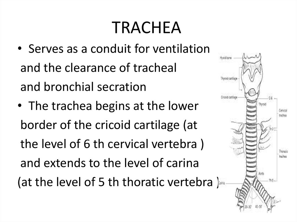
TRACHEA• Serves as a conduit for ventilation
and the clearance of tracheal
and bronchial secration
• The trachea begins at the lower
border of the cricoid cartilage (at
the level of 6 th cervical vertebra )
and extends to the level of carina
(at the level of 5 th thoratic vertebra )
5.
TRACHEA• Length of trachea in averag of 10-13 cm
• and its contain of C shaped cartilage ring
(16-20), witch form the anterior and lateral walls
of trahea , and posteriorly by the membrans
wall ,thes cartilage hold and support the
tracheal and preventing it from coolapsing
but .
6.
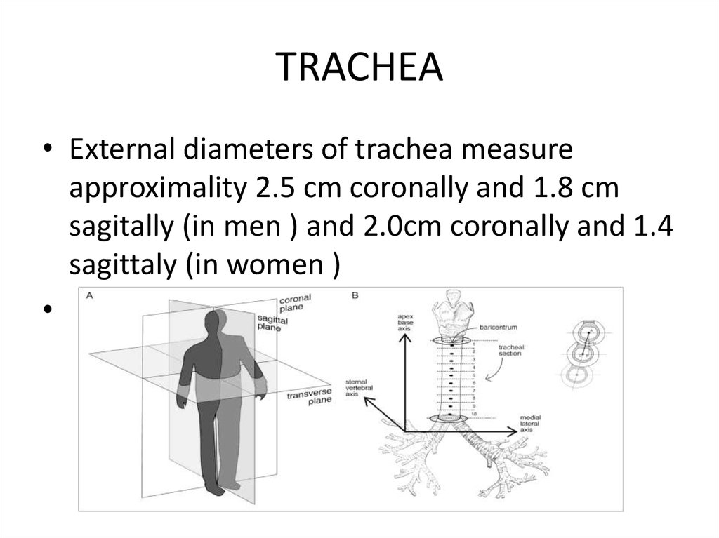
TRACHEA• External diameters of trachea measure
approximality 2.5 cm coronally and 1.8 cm
sagitally (in men ) and 2.0cm coronally and 1.4
sagittaly (in women )
7.
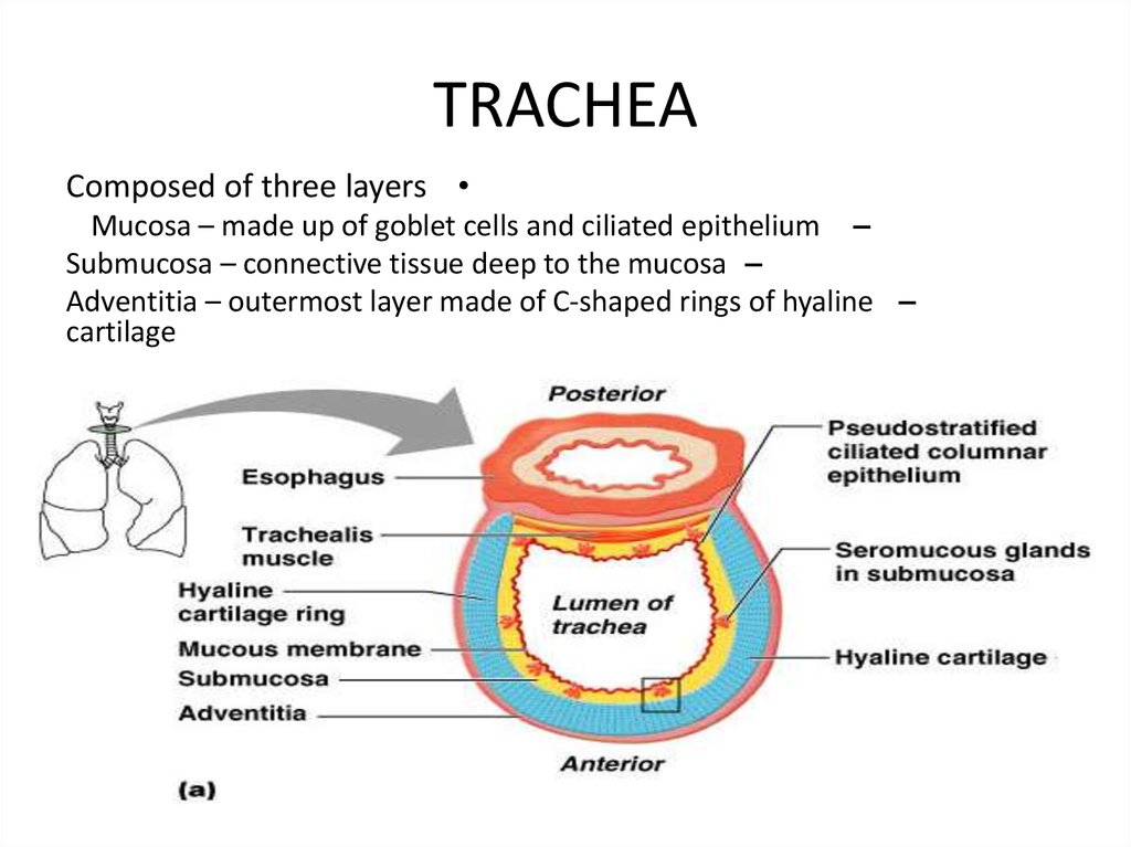
TRACHEAComposed of three layers
Mucosa – made up of goblet cells and ciliated epithelium –
Submucosa – connective tissue deep to the mucosa –
Adventitia – outermost layer made of C-shaped rings of hyaline –
cartilage
8.
BRONCHI• The trachea bifurcates at the level of the
5 th thoratic vertebra ,into the right
and left bronchi
• The right main bronchus is shorter ,
Wider ,and more vertically placed than
the left .
Shorter because it gives off its upper
lobe bronchus sooner (after course
Of only 2.5 cm )
9.
BRONCHI• Wider because it supplies the larger lung
• And vertically (at 25 vetrical compared with 45
on the left ), because the left bronchus has to
extend laterally behind the aortic arch
(inhaled foreign bodeis are moe to enter the
wider and more vertical than narrower )
10.
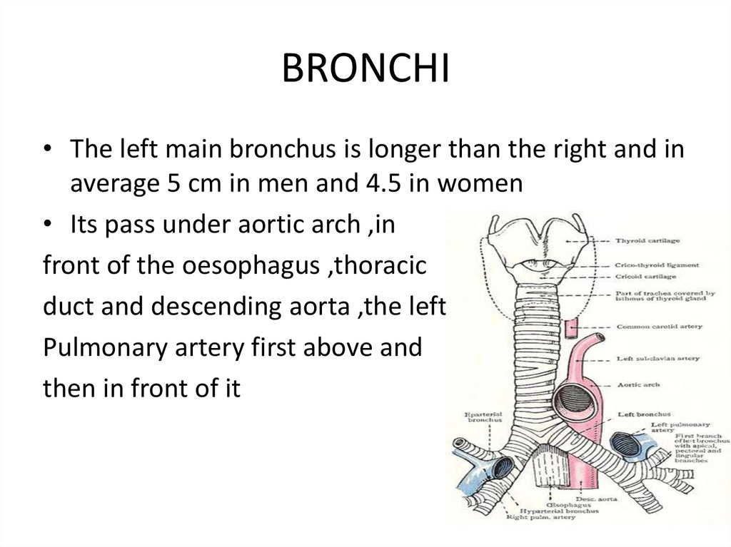
BRONCHI• The left main bronchus is longer than the right and in
average 5 cm in men and 4.5 in women
• Its pass under aortic arch ,in
front of the oesophagus ,thoracic
duct and descending aorta ,the left
Pulmonary artery first above and
then in front of it
11.
The bronchopulmonary• The bronchiols are the finer bronchial ramification ,are usually
of region of 0.6mm in diameter
• The respiratory bronchiols bear small alveoli ,or there walls
and are lined by a nonciliated cuboidal epithelium
• The distal extremity of each respiratpry bronchiole is termed
the alveolar duct
12.
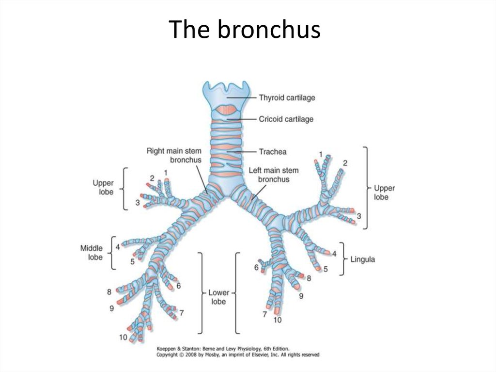
The bronchus• The right lung :
The right main bronchus,after a course of some 2.5 cm , gives off
at right angels the upper lobe bronchus , after 1 cm give
bifurcation into three segmental bronchi 1) apical :upwards
and lateraly 2) posterior :backwards and lateraly 3) anterior
:lateraly and downwards
• The main bronch continues a long 3 cm and give middle lobe
branch , after 1.5 cm give bifurcation into lateral and medial
divisions
• below the middle lobe branchus to apical segment of the
lower lobe ,its 1cm long and gives medial and lateral branches
13.
The bronchus14.
The bronchus• About 1.5 cm below the apical brobchus is given the
medial or cardic bronches , then gives the basal
bronchi : anterior ,lateral,posterior
• The left lung has a course of 5 cm before giveng off
the left upper lobe bronchus ,and thes pass lateraly
for about 1cm and then bifurcates into superior and
inferior , superior supply the apical
• After 1-2cm bifurcated into superior and inferior
15.
ALVEOL• 300 million alveoli in adult for gas exchange
• The alveol a lined with thin and thick side
• In the thin side less than 0.4mic m thick ,where gas exchange
occurs ,the alveolar epithelium and capillary endothelium are
separated only with basement membrane ,
• In the thich side 1-2 thick , where the fluid and solute
exchange occurs,the pulmonary interstitial space( collagen
and nerve fibers ) separates alveolar from capillary
endothelium
16.
ALVEOL• The pulmonary epithelium contains the cells
• A) type 1 pneumocytes :are flat and form 1 –nm junction
with another and thes important to prevent the passage large
active molecules into the alveols
• B) type 2 pneumocytes : are more than type 1 and thes
contain surfactan and cane produce type 1 pneumocytes
17.
The pulmonary blood supply• The blood supply to the lung ,lymphs ,bronchi
is provided by the bronchial arteries
And thes provoids small amount of cardic
output 4%,branch the bronchial artery supply
the bronchi as far as terminal bronch
(anastamosis with pulmonary arterial and
continue to alveolar duct ) below thes level
lung tissue is supporeted by compination the
alveolar gas and pulmonary circulation
18.
Innervation• Sympathic (t2-4) and parasympathic (vagal)
form a posterior pulmonary plexus at the root
of the lung
• Fiber pass around the lung root to form an
anterior pulminary nerve plexus ,from the
plexus to the lung and bronchi